CT image pulmonary nodule detection method, apparatus and device, and readable storage medium

A CT image and detection method technology, applied in the field of image processing, can solve the problems of low detection efficiency and accuracy of artificial pulmonary nodules

Active Publication Date: 2018-12-11
SHENZHEN IMSIGHT MEDICAL TECH CO LTD
View PDF5 Cites 13 Cited by
  • Summary
  • Abstract
  • Description
  • Claims
  • Application Information

AI Technical Summary

Problems solved by technology

[0004] The main purpose of the present invention is to provide a method, device, equipment and readable storage medium for detecting pulmonary

Method used


Image

  • CT image pulmonary nodule detection method, apparatus and device, and readable storage medium
  • CT image pulmonary nodule detection method, apparatus and device, and readable storage medium
  • CT image pulmonary nodule detection method, apparatus and device, and readable storage medium

Examples


Embodiment Construction

[0072] It should be understood that the specific embodiments described here are only used to explain the present invention, not to limit the present invention.

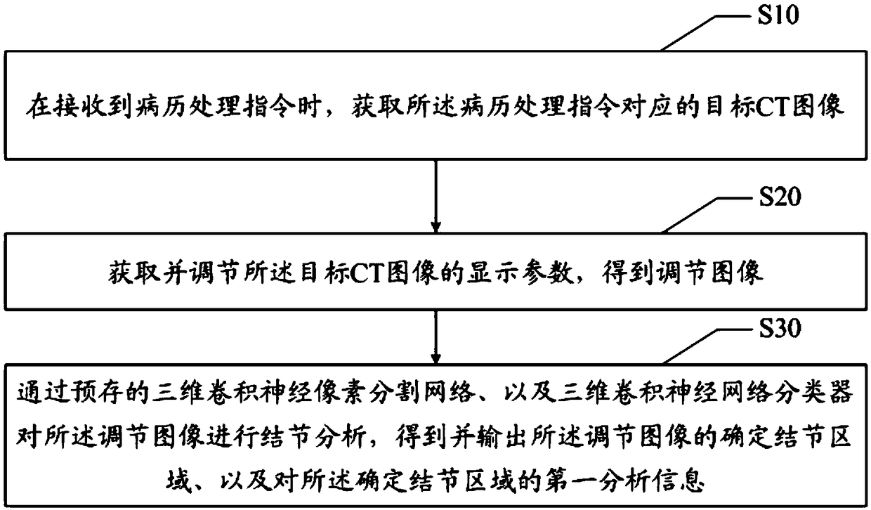
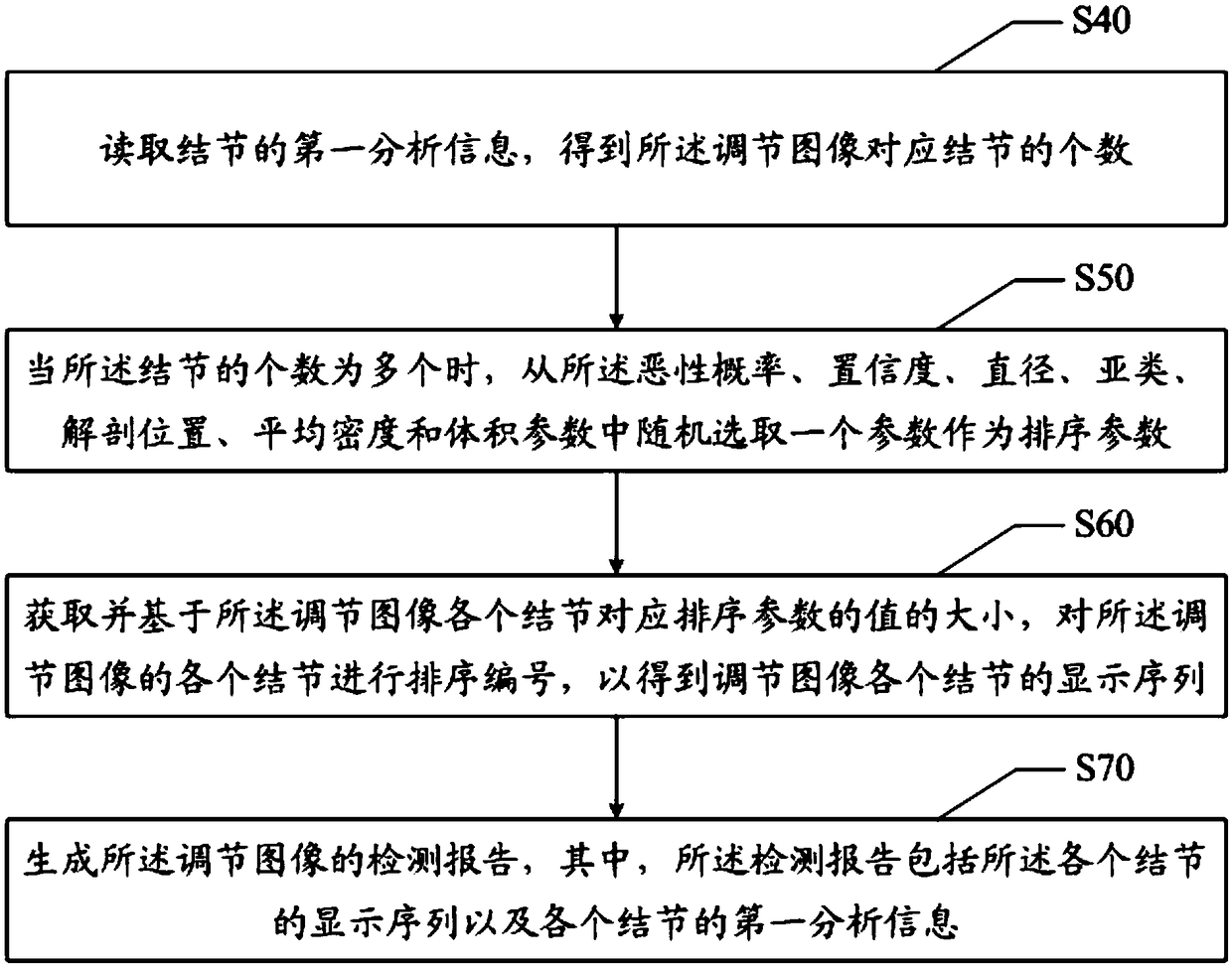
[0073] The present invention provides a method for detecting pulmonary nodules in CT images. In the first embodiment of the method for detecting pulmonary nodules in CT images of the present invention, refer to figure 1 , the CT image pulmonary nodule detection method comprises:

[0074] When a medical record processing instruction is received, the target CT image corresponding to the medical record processing instruction is obtained; the display parameters of the target CT image are obtained and adjusted to obtain an adjusted image; The product neural network classifier analyzes the determined nodule area on the adjustment image, obtains and outputs the determined nodule area of ​​the adjusted image and first analysis information on the determined nodule area.

[0075] Specific steps are as follows:

[0076] Step S...


PUM


Abstract

The invention discloses a CT image lung nodule detection method, apparatus and device and a readable storage medium. The CT image lung nodule detection method comprises the following steps: when receiving a medical record processing instruction, obtaining a target CT image corresponding to the medical record processing instruction; acquiring and adjusting display parameters of the target CT imageto obtain an adjusted image; through the pre-stored three-dimensional convolution neural pixel segmentation network and the three-dimensional convolution neural network classifier, analyzing the adjustment image to determine the nodular region, obtaining and outputting the determined nodular region of the adjustment image, and obtaining the first analysis information of the determined nodular region. The invention solves the technical problems of low detection efficiency and low accuracy of the existing artificial lung nodules.

Description

technical field [0001] The present invention relates to the technical field of image processing, in particular to a method, device, equipment and readable storage medium for detecting pulmonary nodules in CT images. Background technique [0002] Pulmonary nodules are one of the most important early signs of lung cancer, that is, the lesion characteristics of lung lesions can be deduced from the lesion characteristics of lung nodules. [0003] At present, after obtaining the CT images of the lungs, doctors need to manually read the CT images to extract or detect the analysis information of the determined nodule area of ​​the lung. Due to the uncertainty of the size, shape and density of the lung nodules However, artificial pulmonary nodule feature detection is difficult to meet the market's demand for the efficiency and accuracy of pulmonary nodule feature detection. Contents of the invention [0004] The main purpose of the present invention is to provide a method, device...

Claims


Application Information

Patent Timeline
IPC IPC(8): G06T7/00G06N3/04
CPCG06T7/0012G06T2207/30064G06T2207/20081G06T2207/20084G06T2207/10081G06N3/045
Inventor 窦琪刘权德陈浩
Owner SHENZHEN IMSIGHT MEDICAL TECH CO LTD
Why Eureka
Try Eureka