Three-dimensional porous stent and preparation method and application thereof

A three-dimensional porous and porous technology, applied in medical science, prosthesis, additive processing, etc., to achieve precise controllability, good biocompatibility and biodegradability, and suitable pore size

Active Publication Date: 2018-12-28
南京恒华生物发展有限公司
View PDF10 Cites 8 Cited by
  • Summary
  • Abstract
  • Description
  • Claims
  • Application Information

AI Technical Summary

Problems solved by technology

In addition, the scaffolds prepared by this method are only limited to micron-sized spherical or fibrous, which is not suitable for the actual clinical needs of tissue engineering scaffolds with various shapes and sizes.

Method used


Image

  • Three-dimensional porous stent and preparation method and application thereof
  • Three-dimensional porous stent and preparation method and application thereof
  • Three-dimensional porous stent and preparation method and application thereof

Examples


Embodiment 1

[0075] A method for preparing a three-dimensional porous scaffold, comprising:

[0076] 1) Preparation steps of monodisperse emulsion droplets: using a microfluidic device, an external phase and an internal phase to prepare monodisperse emulsion droplets with a controllable particle size, and cleaning and collecting the monodisperse emulsion droplets;

[0077] Wherein, wherein, the external phase includes a composite composed of degradable natural biological materials and degradable synthetic polymer materials; the weight ratio of the degradable natural biological materials to the degradable synthetic polymer materials is 1:7.5 . The degradable natural biological material is sodium alginate. The artificially synthesized polymer material is polyethylene glycol diacrylate.

[0078] The internal phase is methyl silicone oil.

[0079] 2) The preparation steps of the emulsion droplet template: the collected monodisperse emulsion droplets are left to stand, the monodisperse emuls...

Embodiment 2

[0083] 1) Preparation steps of monodisperse emulsion droplets: using a microfluidic device, an external phase and an internal phase to prepare monodisperse emulsion droplets with a controllable particle size, and cleaning and collecting the monodisperse emulsion droplets;

[0084] Wherein, the outer phase 1 includes a composite composed of degradable natural biomaterials and degradable synthetic polymer materials; the weight ratio of the degradable natural biomaterials to the degradable synthetic biomaterials is 1:7.5. The degradable natural biological material is sodium alginate. The synthetic biomaterial is gelatin methacrylate (Gelma).

[0085] The internal phase is methyl silicone oil.

[0086] 2) The preparation steps of the emulsion droplet template: the collected monodisperse emulsion droplets are left to stand, the monodisperse emulsion droplets will self-assemble into a tightly packed structure, and finally form a uniform and neat emulsion droplet template; then the ...

Embodiment 3

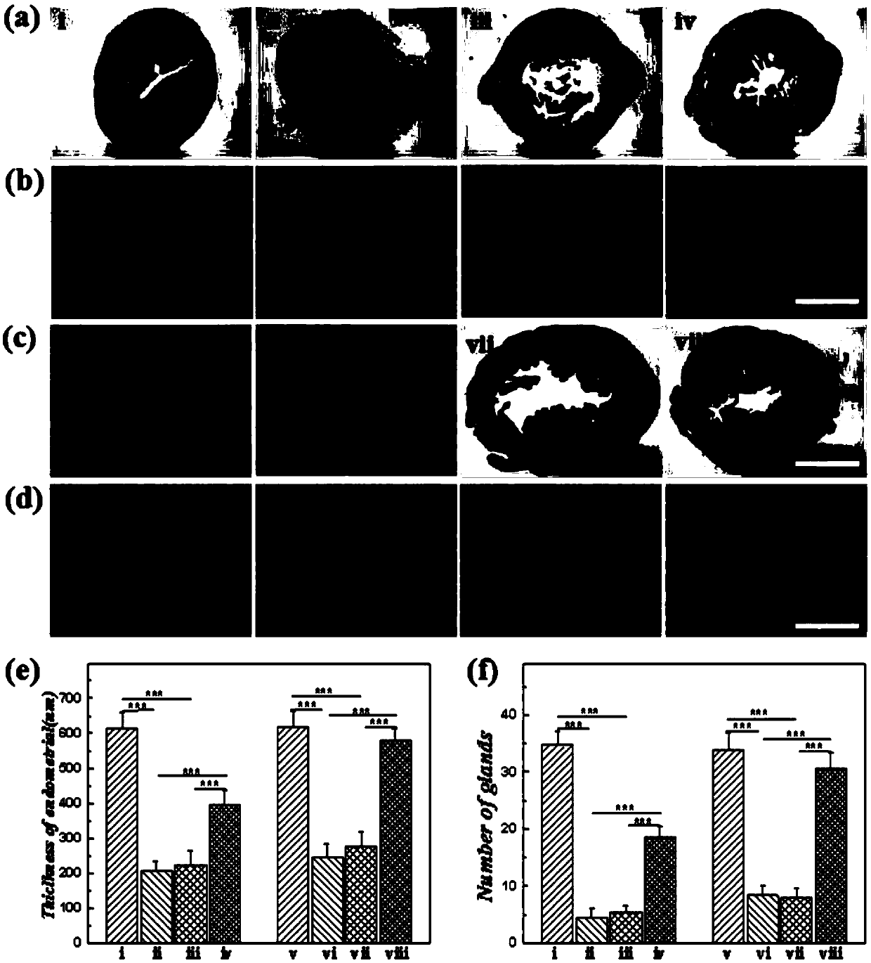
[0091] A method for preparing a drug-loaded three-dimensional porous scaffold: soak the three-dimensional porous scaffold described in Example 3 in a 75% alcohol solution overnight, then wash it repeatedly with sterile PBS (PH=7.4), and soak it in PBS for ultraviolet light Irradiate for 3h. Soak the porous scaffold in 100ug / mL bFGF solution to fully disperse the drug into the pores, take it out after 24 hours for later use, and obtain the drug-loaded three-dimensional porous scaffold.

[0092] Application of drug-loaded three-dimensional porous scaffold in intrauterine adhesion rat model:

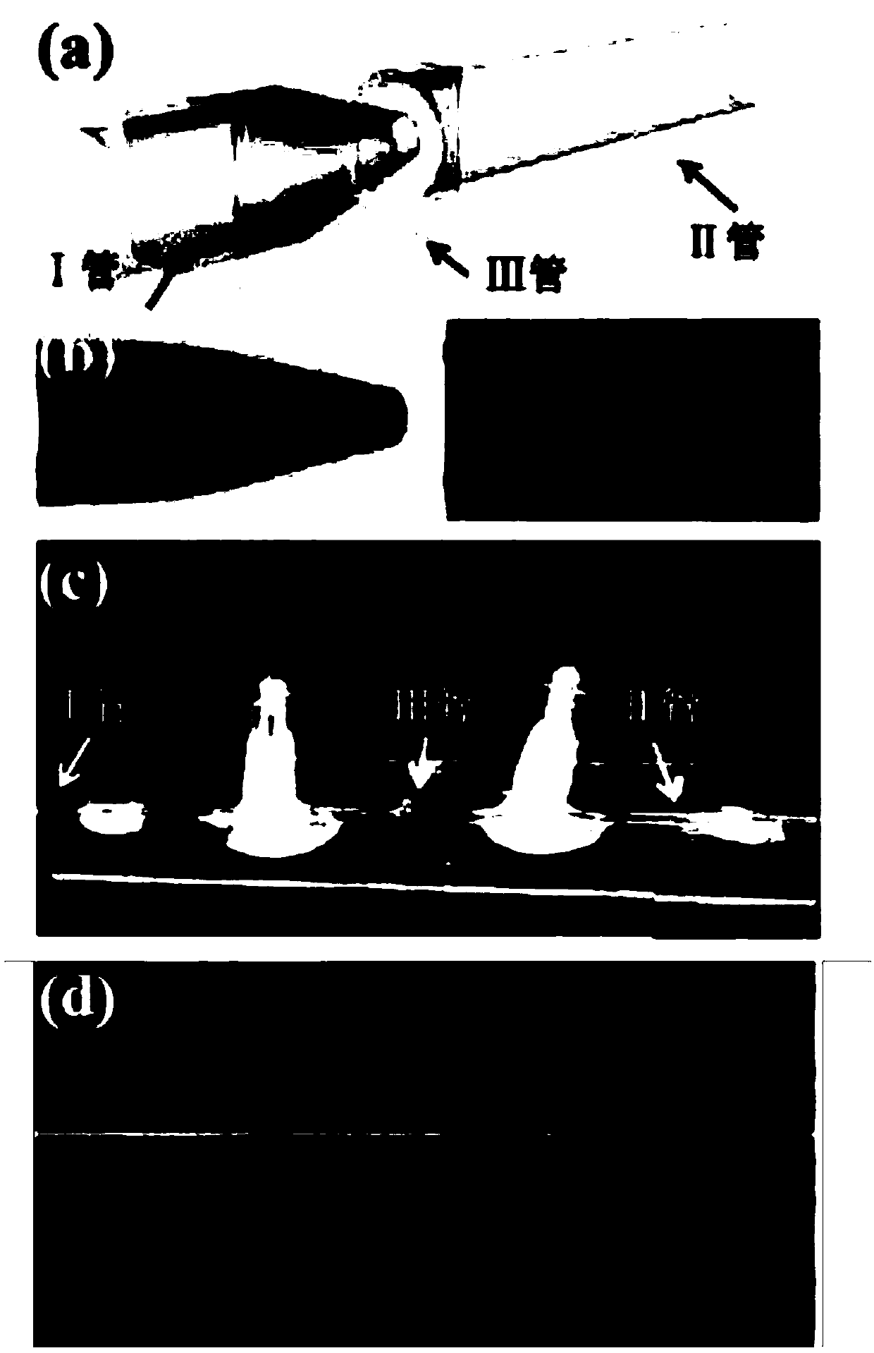
[0093] (1) Establishment of intrauterine adhesion model rats: the rats were anesthetized with 350 mg / kg 10% chloral hydrate, the lower abdomen was shaved and fixed in the supine position on the operating board, disinfected with iodine, and a long slit was made in the middle of the lower abdomen. A longitudinal incision of about 2-3 cm was made to enter the abdominal cavity to expose the ut...


PUM

PropertyMeasurementUnit
pore sizeaaaaaaaaaa
pore sizeaaaaaaaaaa

Abstract

The invention discloses a three-dimensional porous stent. The three-dimensional porous stent is made from composite biomaterials consists of degradable natural biomaterials and degradable artificiallysynthesized macromolecule materials. The three-dimensional porous stent has a three-dimensional orderly porous structure in which holes are homogeneous, mutually penetrated and regular in distribution, and the hole diameter is 80-350[mu]m. The invention further discloses a preparation method and application of the three-dimensional porous stent. According to the method disclosed by the invention,the advantages of an emulsion microflow control technique and the advantages of the composite biomaterials are combined. The three-dimensional porous stent disclosed by the invention has favorable biocompatibility and biodegradability and has the characteristics that the hole diameter is appropriate, the holes are mutually penetrated, and the three-dimensional porous stent is degradable and compressible, can be shaped and can be used for performing drainage effectively, and good space is provided for medicine delivery and slow release, cell growth and metabolism and tissue repair; the proportion of ingredients of the compound materials and the holes in concentrated distribution can guarantee that the three-dimensional porous stent has favorable compression and resilience properties like sponge, so that the three-dimensional porous stent can better adapt to the shape of the uterine cavity or the shape of other positions needing prevention of adhesion, and the effect of effectively preventing the adhesion can be achieved.

Description

technical field [0001] The invention relates to the technical field of three-dimensional porous tissue engineering scaffold materials, in particular to a three-dimensional porous scaffold and its preparation method and application. Background technique [0002] Intrauterine adhesions (IUA), also known as Asherman syndrome, are caused by damage to the basal layer of the endometrium caused by various reasons, resulting in partial or complete adhesions of the cervix or uterine cavity. Its clinical manifestations are irregular menstruation, secondary infertility, repeated premature delivery, miscarriage, other adverse pregnancy outcomes and even fetal death, which seriously affect women's physical and mental health. Although transcervical adhesion resection (TCRA) is currently the main method for the treatment of intrauterine adhesions, the recurrence rate of postoperative adhesions is still high, and the recurrence rate of severe intrauterine adhesions is as high as 60%. There...

Claims


Application Information

Patent Timeline
Patent Type & Authority Applications(China)
IPC IPC(8): A61L27/56A61L27/50A61L27/54A61L27/22A61L27/38B33Y10/00B33Y80/00
CPCA61L27/227A61L27/3804A61L27/50A61L27/54A61L27/56A61L2300/412A61L2300/414A61L2300/602B33Y10/00B33Y80/00C08L89/00
Inventor 吕洋周晓萍蔡云朗吴方媛余筠茹
Owner 南京恒华生物发展有限公司
Why Eureka
Try Eureka