
Fully-automatic pelvic tumor segmentation method and system, storage medium and terminal

A fully automatic, oncological technology, applied in the field of medical image processing, can solve the problems of low repeatability, difficulty in obtaining a large number of segmentation results, time-consuming and labor-intensive problems, etc., to reduce workload, shorten medical-industrial interaction time, and achieve high accuracy Effect

Active Publication Date: 2019-01-15
SHANGHAI NINTH PEOPLES HOSPITAL AFFILIATED TO SHANGHAI JIAO TONG UNIV SCHOOL OF MEDICINE
View PDF4 Cites 6 Cited by
  • Summary
  • Abstract
  • Description
  • Claims
  • Application Information

AI Technical Summary

Problems solved by technology

[0004] (1) Time-consuming and labor-intensive, it is difficult to quickly obtain a large number of segmentation results;
[0

Method used


Image

  • Fully-automatic pelvic tumor segmentation method and system, storage medium and terminal
  • Fully-automatic pelvic tumor segmentation method and system, storage medium and terminal
  • Fully-automatic pelvic tumor segmentation method and system, storage medium and terminal

Examples


Embodiment Construction

[0042] Embodiments of the present invention are described below through specific examples, and those skilled in the art can easily understand other advantages and effects of the present invention from the content disclosed in this specification. The present invention can also be implemented or applied through other different specific implementation modes, and various modifications or changes can be made to the details in this specification based on different viewpoints and applications without departing from the spirit of the present invention. It should be noted that, in the case of no conflict, the following embodiments and features in the embodiments can be combined with each other.

[0043] It should be noted that the diagrams provided in the following embodiments are only schematically illustrating the basic ideas of the present invention, and only the components related to the present invention are shown in the diagrams rather than the number, shape and number of componen...


PUM


Abstract

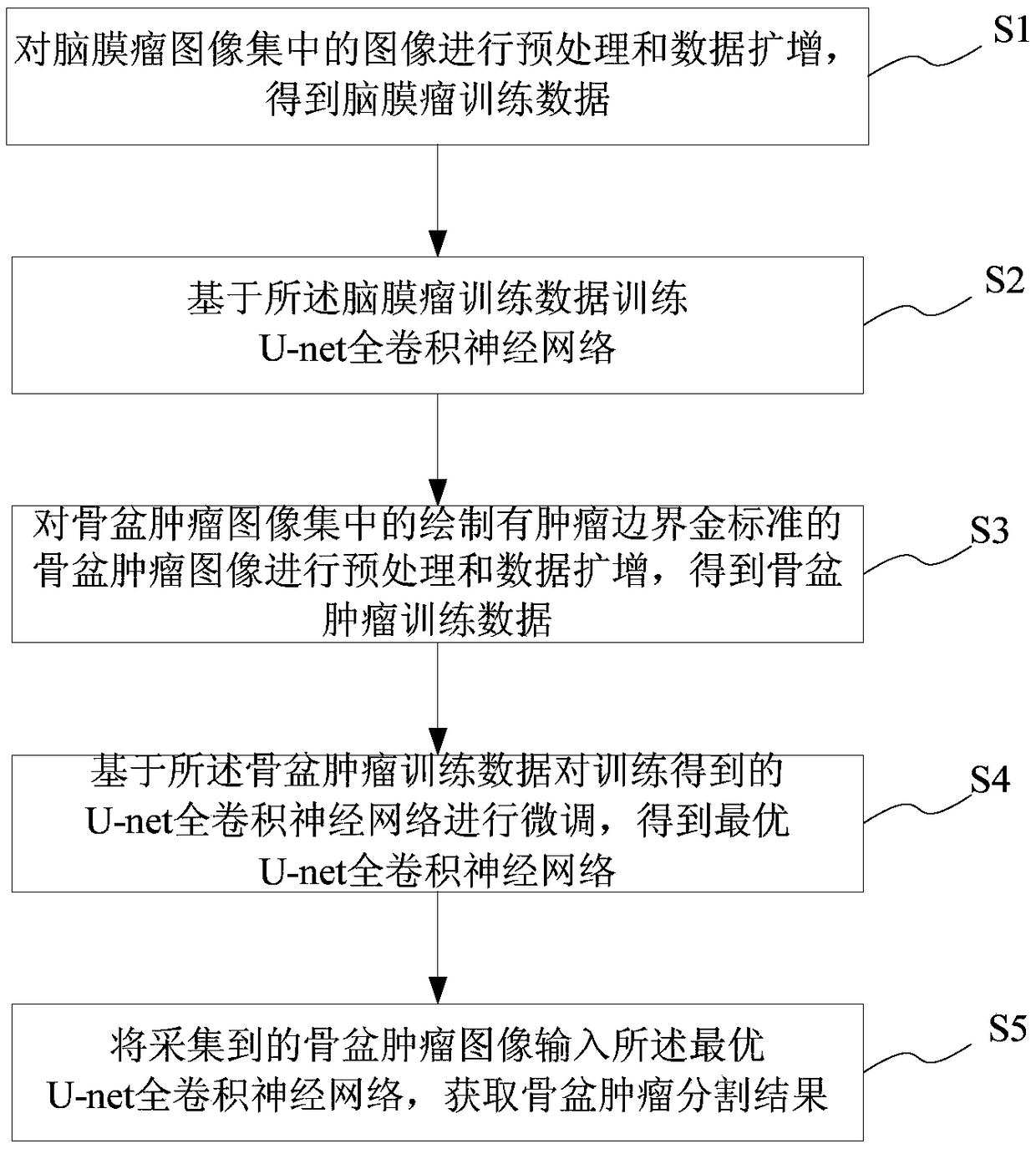
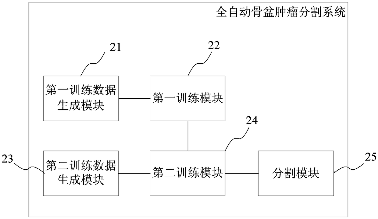
The invention provides a full-automatic pelvic tumor segmentation method and system, a storage medium and a terminal thereof, comprising the following steps: preprocessing and data amplification are carried out on images in a meningioma image set to obtain meningioma training data; training U-net convolutional neural network based on the meningioma training data; the pelvic tumor training data were obtained by preprocessing and data amplification of the pelvic tumor images with tumor boundary gold standard drawn in the pelvic tumor image set. Training data of the pelvic tumor based on the training results of the U-net to realize fine-tuning of the full convolution neural network to get the optimal U-net fully convolution neural networks; inputting the acquired pelvic tumor image into the optimal U-Net total convolution neural network to obtain the segmentation results of pelvic tumors. The invention discloses a full-automatic pelvic tumor segmentation method and system, a storage medium and a terminal through migration learning U-net fully convolutional neural network to realize automatic pelvic tumor segmentation, which greatly improves the efficiency of medical interaction and the accuracy of pelvic tumor segmentation.

Description

technical field [0001] The present invention relates to the technical field of medical image processing, in particular to a fully automatic pelvic tumor segmentation method and system, a storage medium and a terminal. Background technique [0002] Among tumors of the musculoskeletal system, there is usually significant heterogeneity in both primary and secondary bone tumors. Especially for complex pelvic tumors, the lesions involve multiple pelvic partitions, and the surgical resection range is large, making the automatic segmentation of pelvic tumors always a big challenge. [0003] At present, tumor boundary image judgment is the link with the lowest efficiency and the most impact on surgical results in medical-industrial interaction engineering. The existing pelvic tumor segmentation methods are usually marked and segmented manually by experienced physicians, which have the following defects: [0004] (1) Time-consuming and labor-intensive, it is difficult to quickly ob...

Claims


Application Information

Patent Timeline
IPC IPC(8): G06T7/12G06T7/30G06N3/04
CPCG06T7/12G06T7/30G06T2207/20084G06T2207/20081G06T2207/10088G06T2207/30096G06T2207/30016G06N3/045
Inventor 艾松涛王燎曲扬李小敏
Owner SHANGHAI NINTH PEOPLES HOSPITAL AFFILIATED TO SHANGHAI JIAO TONG UNIV SCHOOL OF MEDICINE
Learn More