A medical image processing method, system and apparatus, and computer-readable storage medium

An image processing and medical image technology, applied in the field of medical image processing, can solve the problems that the segmentation performance cannot meet the clinical needs, the tumor boundary is unclear, and the segmentation accuracy is low, so as to achieve a good segmentation effect, good segmentation results, and improve the The effect of segmentation accuracy

Inactive Publication Date: 2019-01-25
SHANGHAI UNITED IMAGING INTELLIGENT MEDICAL TECH CO LTD
View PDF4 Cites 27 Cited by
  • Summary
  • Abstract
  • Description
  • Claims
  • Application Information

AI Technical Summary

Problems solved by technology

In addition, due to tumor heterogeneity, the tumor boundary is unclear and the segmentation accuracy is low
Although the current deep learning method can automatically extract features, the segmentation performance and segmentation speed still cannot meet the clinical needs

Method used


Image

  • A medical image processing method, system and apparatus, and computer-readable storage medium
  • A medical image processing method, system and apparatus, and computer-readable storage medium
  • A medical image processing method, system and apparatus, and computer-readable storage medium

Examples


Embodiment Construction

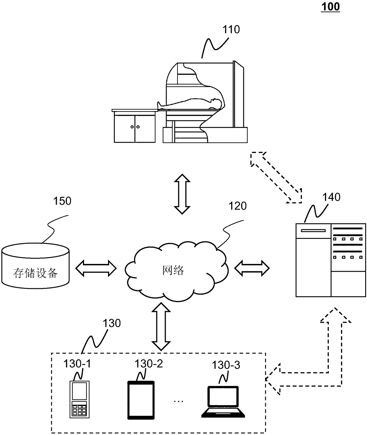
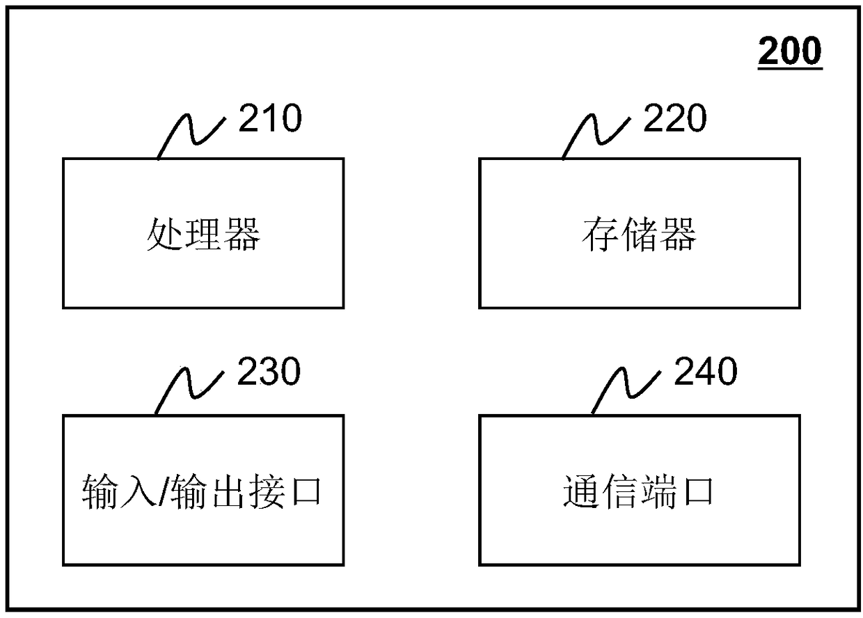
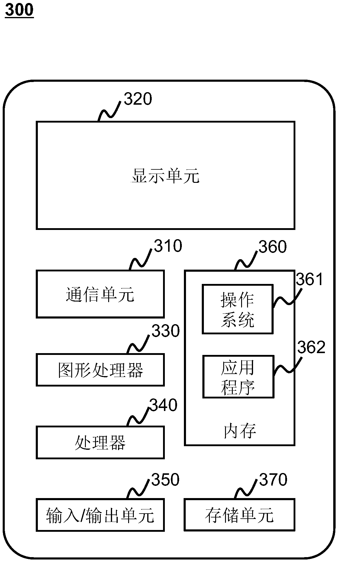
[0025] In order to more clearly illustrate the technical solutions of the embodiments of the present application, the following briefly introduces the drawings that need to be used in the description of the embodiments. Illustrative diagrams Obviously, the accompanying drawings in the following description are only some examples or embodiments of the present application, and those of ordinary skill in the art can also apply the present application according to these drawings without creative work. applied to other similar situations. Unless otherwise apparent from context or otherwise indicated, like reference numerals in the figures represent like structures or operations.

[0026] It should be understood that "system", "device", "unit" and / or "module" as used herein is a method used to distinguish different components, elements, parts, parts or assemblies of different levels. However, the words may be replaced by other expressions if other words can achieve the same purpose...


PUM


Abstract

Embodiments of the present application disclose a medical image processing method, system and apparatus, and computer-readable storage medium. The method comprises the following steps: acquiring a sample image, wherein the sample image comprises an abnormal region, the abnormal region comprises at least one sub-abnormal region; Acquiring an image processing cascade network, the image processing cascade network comprising at least two levels of neural networks; Training the neural networks in the image processing cascade network with the sample images to obtain a fully trained image processingcascade network; Wherein the input of the first-level neural network is an unsegmented sample image, and the output of the last-level neural network is a segmentation result of the at least one sub-abnormal region; The trained image processing cascade network is used to process the image to be tested and determine the abnormal region in the image to be tested.

Description

technical field [0001] The present application relates to the field of medical image processing, in particular to a method and system for medical image segmentation. Background technique [0002] Most of the existing medical image processing software only processes images of organs or tissues with normal appearance, and it is difficult to process images of organs or tissues with lesions or injuries. Taking brain images as an example, existing medical image processing software can handle normal brain images well, but for abnormal brain images such as brain tumors, strokes, cerebral hemorrhages, and multiple sclerosis, existing There are many deficiencies in the processing of medical image processing software. Taking brain tumors as an example, due to the existence of tumor heterogeneity, the interior of brain tumors can be divided into peritumoral edema, tumor core, and tumor enhancement area. In the formulation of the surgical plan, the tumor core usually needs to be compl...

Claims


Application Information

Patent Timeline
Patent Type & Authority Applications(China)
IPC IPC(8): G06K9/34G06N3/04
CPCG06V10/267G06V2201/03G06N3/045
Inventor 花蕊石峰高耀宗詹翊强
Owner SHANGHAI UNITED IMAGING INTELLIGENT MEDICAL TECH CO LTD
Why Eureka
Try Eureka