A Brain Tissue Extraction Method Based on Fully Convolutional Neural Network
A technology of convolutional neural network and extraction method, applied in the field of digital image processing, can solve the problem of difficult accurate segmentation of brain tissue, achieve the effect of refined segmentation goals and ensure computing efficiency
Active Publication Date: 2021-11-02
SOUTHEAST UNIV
View PDF3 Cites 0 Cited by
- Summary
- Abstract
- Description
- Claims
- Application Information
AI Technical Summary
Problems solved by technology
At present, the existing traditional brain tissue extraction methods are difficult to achieve accurate segmentation of brain tissue
Method used
View moreImage
Smart ImageViewing Examples

Examples
Embodiment
[0061] The brain tissue extraction method based on the fully convolutional neural network of the present invention will be described below by taking the OASIS data set, the IBSR data set and the LPBA40 data set as examples respectively.
Login to view more PUM
 Abstract
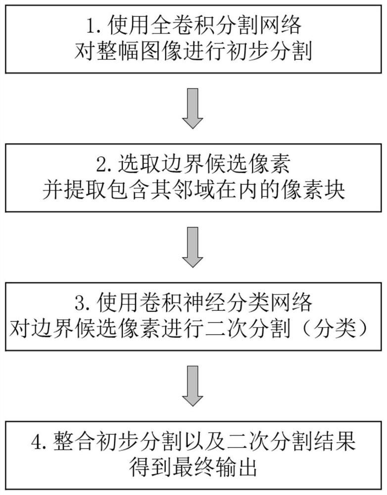
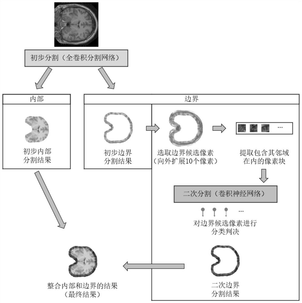
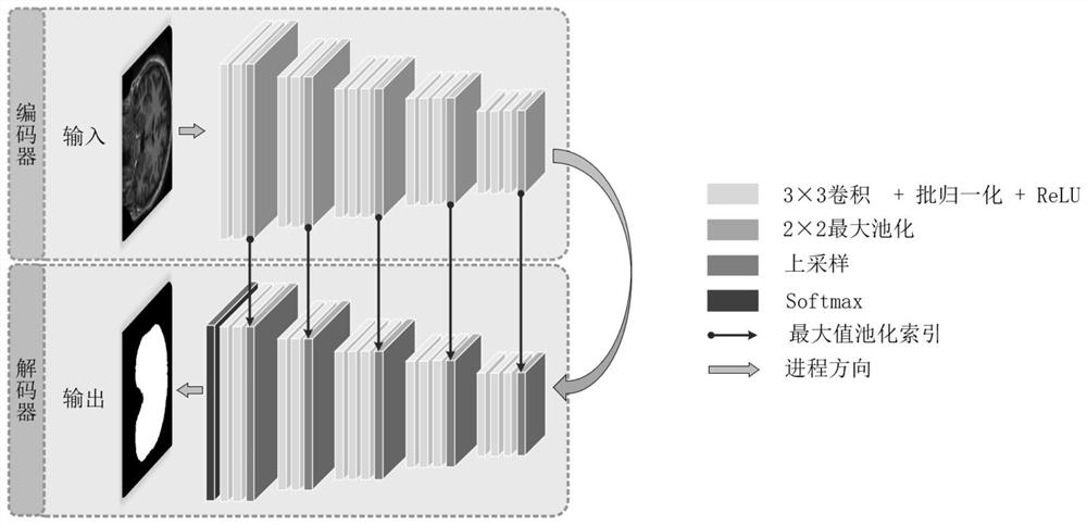
The invention discloses a method for extracting brain tissue based on a fully convolutional neural network, comprising the following steps: firstly, using a fully convolutional segmentation network to perform preliminary segmentation on a two-dimensional original nuclear magnetic resonance image to obtain a preliminary segmentation result; secondly, according to The preliminary segmentation results separate the internal and boundary information of the brain tissue; again, select these pixels that cannot be determined to be brain tissue as boundary candidate pixels, and send these candidate pixels and their neighborhoods to the convolutional neural network for secondary segmentation. Realize the classification judgment; finally, integrate the internal segmentation results obtained from the primary segmentation and the boundary segmentation results obtained from the secondary segmentation, and then obtain the final segmentation results of brain tissue extraction. The present invention performs two thick and thin segmentations, which not only ensures the calculation efficiency of the method, but also realizes the refined segmentation target, and can be better applied to brain magnetic resonance images to achieve more accurate brain tissue and skull, eyeballs, skin, The removal of non-brain tissue such as fat.
Description
technical field [0001] The invention belongs to the technical field of digital image processing, and relates to a brain tissue extraction method based on a fully convolutional neural network. Background technique [0002] The brain is one of the vital organs of the human body and an important part of our body. Human beings have never stopped studying the brain. Scientists hope to explore the unknown functions of the human brain by studying the complex structure inside the brain. Magnetic Resonance Imaging (MRI) technology is non-invasive, contains a large amount of information, and has the characteristics of multi-directional imaging. In MRI images, soft tissues with relatively low gray scale can be clearly distinguished. Therefore, important information such as the location and size of brain tissue anatomy in MRI images can be identified with the naked eye. In addition, MRI images have been widely used clinically because of their high signal-to-noise ratio, high resoluti...
Claims
Login to view more Application Information
Patent Timeline
 Patent Type & Authority Patents(China)
IPC IPC(8): G06T7/00G06T7/10
CPCG06T7/0012G06T2207/10088G06T2207/20084G06T2207/30016G06T7/10
Inventor 舒华忠吴颖真赵仕进孔佑勇
Owner SOUTHEAST UNIV
- R&D Engineer
- R&D Manager
- IP Professional
Why Eureka
- Industry Leading Data Capabilities
- Powerful AI technology
- Patent DNA Extraction
Try Eureka
Browse by: Latest US Patents, China's latest patents, Technical Efficacy Thesaurus, Application Domain, Technology Topic.
© 2024 PatSnap. All rights reserved.Legal|Privacy policy|Modern Slavery Act Transparency Statement|Sitemap