A Multi-Scale Imaging Method for Ultrasonic Scattering Sub-diameters Based on Backscattering Coefficient

An imaging method and scatterer technology, applied in the field of signal processing, can solve the problems of missing frequency and limited diagnostic information of ultrasonic imaging.

Active Publication Date: 2021-06-18
BEIJING UNIV OF TECH
View PDF7 Cites 0 Cited by
  • Summary
  • Abstract
  • Description
  • Claims
  • Application Information

AI Technical Summary

Problems solved by technology

The commonly used clinical "B-ultrasound" (B-mode ultrasound imaging) uses the amplitude information of the backscattered signal (radio frequency signal) for imaging, but loses information such as frequency, resulting in limited diagnostic information of ultrasound imaging

Method used


Image

  • A Multi-Scale Imaging Method for Ultrasonic Scattering Sub-diameters Based on Backscattering Coefficient
  • A Multi-Scale Imaging Method for Ultrasonic Scattering Sub-diameters Based on Backscattering Coefficient
  • A Multi-Scale Imaging Method for Ultrasonic Scattering Sub-diameters Based on Backscattering Coefficient

Examples


Embodiment Construction

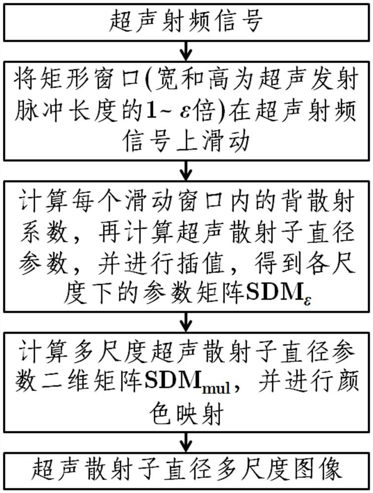
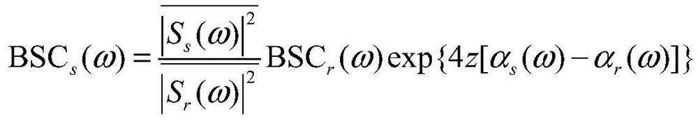
[0028] The multi-scale imaging method of ultrasonic scatterer diameter based on backscatter coefficient of the present invention is based on the ultrasonic backscatter signal (radio frequency signal) of the tissue to be measured, calculates the backscatter coefficient, and then calculates the ultrasonic scatterer diameter parameter and calculates the ultrasonic scatterer diameter Parametric methods for multiscale images.

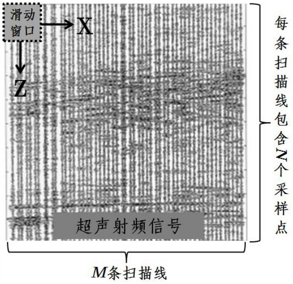
[0029] Without loss of generality, it is assumed that the ultrasonic radio frequency signal is composed of M scanning lines, each scanning line contains N sampling points, and the distance between two adjacent scanning lines is Int lat (unit is meter), the distance between two adjacent sampling points is Int axi (the unit is meter), then the ultrasonic radio frequency signal is a two-dimensional matrix with a size of M×N; the length of the ultrasonic emission pulse is set to be Len (the unit is meter). figure 1 Be the flowchart of the inventive method, main...


PUM


Abstract

The invention discloses a multi-scale imaging method of ultrasonic scatterer diameters based on backscatter coefficients, which is based on ultrasonic radio frequency signals, calculating backscatter coefficients, calculating parameters of ultrasonic scatterer diameters, and calculating multi-scale images of ultrasonic scatterer diameters. Slide the sliding windows of different scales on the ultrasonic radio frequency signal, and calculate the ultrasonic scattering sub-diameter parameters in each sliding window based on the backscattering coefficient, and obtain the ultrasonic scattering sub-diameter parameter value matrix at each scale. The value matrix of the scattering sub-parameters is interpolated to the size of the ultrasonic radio frequency signal, and superimposed and averaged to obtain a multi-scale ultrasonic scattering sub-diameter parameter matrix, and the multi-scale ultrasonic scattering sub-diameter parameter matrix is ​​color-mapped to obtain a multi-scale image of the ultrasonic scattering sub-diameter. The ultrasonic scattering sub-diameter multi-scale imaging method of the present invention can be used for ultrasonic tissue characterization of breast, liver and other biological tissues.

Description

technical field [0001] The invention belongs to the technical field of signal processing, and in particular relates to a medical ultrasonic signal processing method. More specifically, it uses ultrasonic backscatter signals (radio frequency signals) to calculate backscatter coefficients, and then calculates ultrasonic scattering sub-diameter parameters and performs multi-scale imaging. method. Background technique [0002] Ultrasound imaging is widely used in clinical diagnosis due to its real-time performance, low cost, and no ionizing radiation. The ultrasonic probe (transducer) emits ultrasonic waves into the tissue, and after a series of interactions between the ultrasonic waves and the tissue such as scattering, reflection, and diffraction, the ultrasonic probe receives the backscattered echoes of the tissue. The commonly used clinical "B-ultrasound" (B-mode ultrasound imaging) uses the amplitude information of the backscattered signal (radio frequency signal) for imag...

Claims


Application Information

Patent Timeline
Patent Type & Authority Patents(China)
IPC IPC(8): A61B8/00
CPCA61B8/52
Inventor 周著黄欧阳亚丽吴水才高宏建丁琪瑛
Owner BEIJING UNIV OF TECH
Why Eureka
Try Eureka