Magnetic resonance imaging method and system
A magnetic resonance imaging and imaging sequence technology, which is applied in the direction of using nuclear magnetic resonance imaging system for measurement, magnetic resonance measurement, and magnetic variable measurement, can solve the problems of long time consumption of cardiac images and low imaging efficiency, and reduce data acquisition. Time-consuming, improving imaging efficiency, and reducing the number of acquisitions
- Summary
- Abstract
- Description
- Claims
- Application Information
AI Technical Summary
Problems solved by technology
Method used
Image
Examples
Embodiment 1
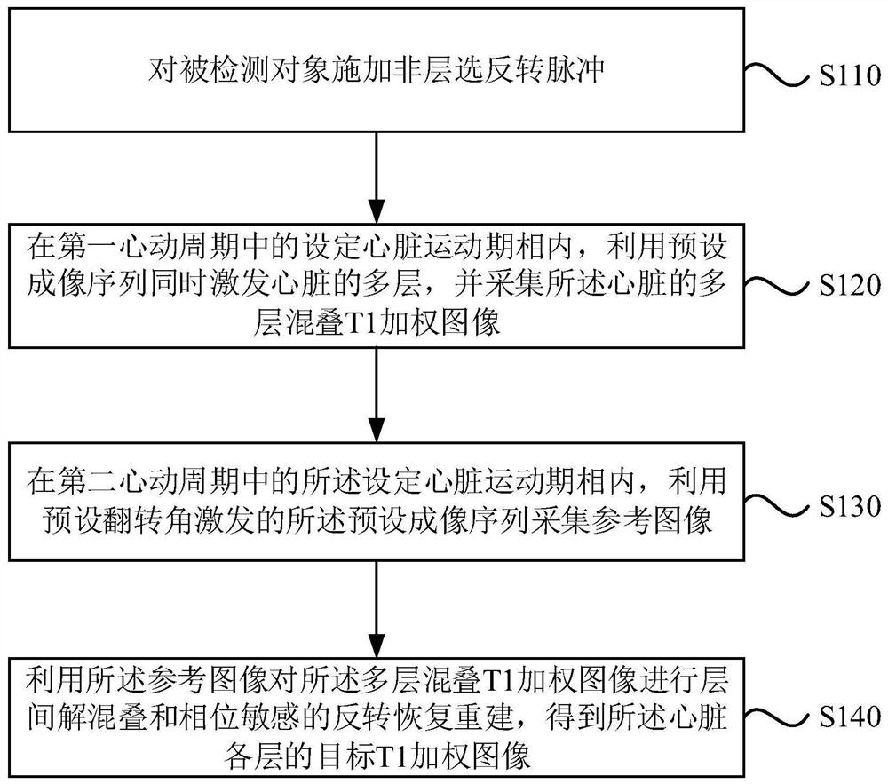
[0029] The magnetic resonance imaging method provided in this embodiment is applicable to fast magnetic resonance imaging of the heart. The method can be performed by a magnetic resonance imaging device, the device can be realized by software and / or hardware, and the device can be integrated in a medical device that can perform magnetic resonance imaging. see figure 1 , the method of this embodiment specifically includes the following steps:
[0030] S110, applying a non-layer selection inversion pulse to the detected object.
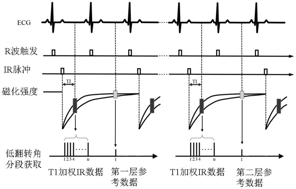
[0031] Specifically, in the process of magnetic resonance imaging of the heart, in order to obtain a stable and effective heart image, the automaticity of the heart movement is used to select the timing of data acquisition. During specific implementation, the extraction is in the static magnetic field B 0 The segmented electrocardiograph (ECG) of the detected object is used as a trigger signal, and a non-layer-selected inversion pulse is applied to t...
Embodiment 2
[0050] In this embodiment, on the basis of the first embodiment above, "use the reference image to perform interlayer de-aliasing and phase-sensitive inversion recovery reconstruction on the multi-layer aliased T1-weighted image to obtain the The target T1-weighted image" was further optimized. The explanations of terms that are the same as or corresponding to the above-mentioned embodiments will not be repeated here. see image 3 , the magnetic resonance imaging method provided in this embodiment includes:
[0051] S310, applying a non-layer selection inversion pulse to the detected object.
[0052] S320. In a set cardiac motion phase in the first cardiac cycle, use a preset imaging sequence to simultaneously excite multiple layers of the heart, and acquire an aliased T1-weighted image of the multiple layers of the heart.
[0053] S330. In the set cardiac motion phase in the second cardiac cycle, use the preset imaging sequence excited by a preset flip angle to acquire a r...
Embodiment 3
[0068] In this embodiment, on the basis of the foregoing embodiments, the "multi-layer aliased T1-weighted image" is further limited. On this basis, further carry out "using the reference image to perform interlayer de-aliasing and phase-sensitive inversion recovery reconstruction on the multi-layer aliased T1-weighted image to obtain the target T1-weighted image of each layer of the heart" Optimized. The explanations of terms that are the same as or corresponding to the above-mentioned embodiments will not be repeated here. see Figure 4 , the magnetic resonance imaging method provided in this embodiment includes:
[0069] S410, applying a non-layer selection inversion pulse to the detected object.
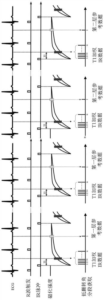
[0070] S420. In the set cardiac motion phase in the first cardiac cycle, use a preset imaging sequence to simultaneously excite multiple layers of the heart, and acquire a multi-layer aliased T1-weighted image of the heart, the multi-layer aliased T1 The K-space corresponding...
PUM
 Abstract
Description
Claims
Application Information
 - R&D
- Intellectual Property
- Life Sciences
- Materials
- Tech Scout
- Unparalleled Data Quality
- Higher Quality Content
- 60% Fewer Hallucinations
Browse by: Latest US Patents, China's latest patents, Technical Efficacy Thesaurus, Application Domain, Technology Topic, Popular Technical Reports.
© 2025 PatSnap. All rights reserved.Legal|Privacy policy|Modern Slavery Act Transparency Statement|Sitemap|About US| Contact US: help@patsnap.com


