Image segmentation method, device, diagnosis system and storage medium

An image segmentation and image technology, applied in the computer field, can solve the problem of poor tumor image segmentation effect and so on

Active Publication Date: 2019-12-27
TENCENT TECH (SHENZHEN) CO LTD +1
View PDF7 Cites 1 Cited by
  • Summary
  • Abstract
  • Description
  • Claims
  • Application Information

AI Technical Summary

Problems solved by technology

[0006] In order to solve the problem of poor tumor image segmentation in the related art, each embodiment of the present invention provides an image segmentation method, device, diagnostic system and storage medium

Method used


Image

  • Image segmentation method, device, diagnosis system and storage medium
  • Image segmentation method, device, diagnosis system and storage medium
  • Image segmentation method, device, diagnosis system and storage medium

Examples


Embodiment Construction

[0036] Here, an exemplary embodiment will be described in detail, and examples thereof are shown in the accompanying drawings. When the following description refers to the accompanying drawings, unless otherwise indicated, the same numbers in different drawings represent the same or similar elements. The implementation manners described in the following exemplary embodiments do not represent all implementation manners consistent with the present invention. Rather, they are merely examples of devices and methods consistent with some aspects of the present invention as detailed in the appended claims.

[0037] As mentioned earlier, tumor image segmentation is mainly based on deep learning, including fully convolutional neural network methods and U-net-based network methods.

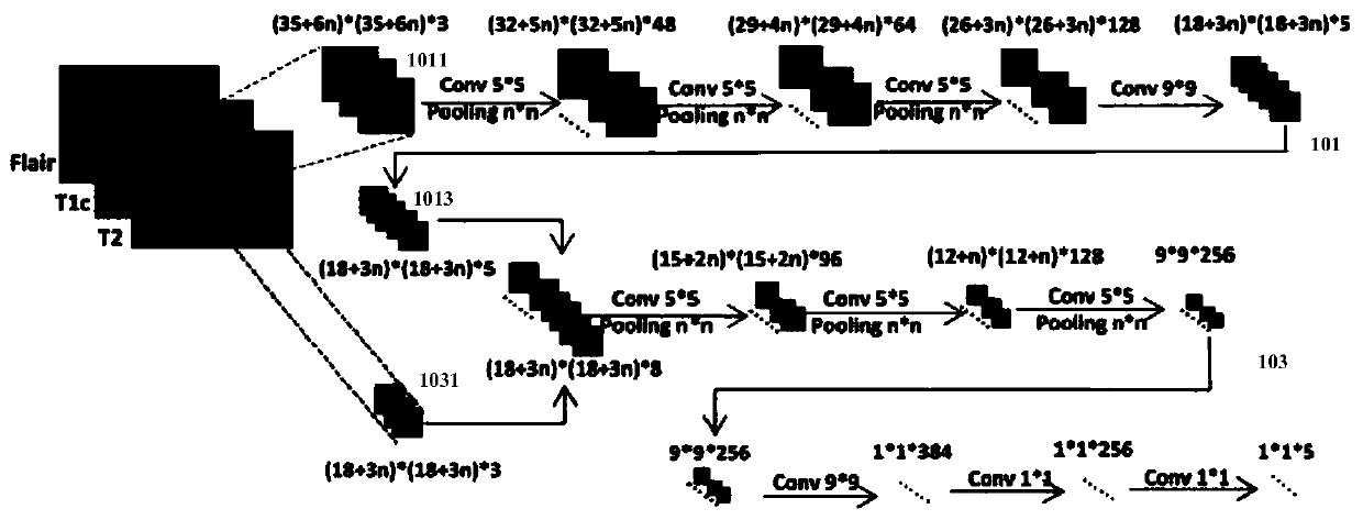
[0038] Among them, such as figure 1 As shown, the fully convolutional neural network method uses two parallel branches 101 and 103 to perform feature learning on images 1011 and 1031 of different sizes. A large...


PUM


Abstract

The invention discloses an image segmentation method, device, diagnosis system and storage medium. The image segmentation method includes: acquiring a tumor image; performing tumor location on the acquired tumor image, and obtaining an image used to indicate all tumors in the tumor image. A candidate image of the region position; the candidate image is input to a cascade segmentation network constructed based on a machine learning model; the first-level segmentation network in the cascade segmentation network is used to perform the full tumor region in the candidate image Image segmentation is the starting point, step by step to the last level of segmentation network to perform image segmentation on the enhanced tumor core area to obtain segmented images. The image segmentation method, device, diagnosis system and storage medium provided by the present invention solve the problem of poor tumor image segmentation effect in the prior art.

Description

Technical field [0001] The present invention relates to the field of computer technology, and in particular to an image segmentation method, device, diagnosis system and storage medium. Background technique [0002] Gliomas are the most common primary malignant brain tumors, also known as brain tumors, with varying degrees of invasiveness, and are often divided into whole tumor areas, tumor core areas, and enhanced tumor core areas. [0003] Magnetic resonance imaging (Magnetic Resonance Imaging, MRI) is the most commonly used method for brain tumor inspection and diagnosis in clinical practice. It can accurately segment the brain tumors from the images generated by multi-modal MRI scans. Medical value. [0004] Currently, tumor image segmentation is mainly based on deep learning, including Fullyconvolutional Neural Networks (FCNNS) method and U-net based network method. [0005] However, the inventor found that the features learned by the full convolutional neural network method are...

Claims


Application Information

Patent Timeline
Patent Type & Authority Patents(China)
IPC IPC(8): G06T7/11G06N99/00
CPCG06T7/11G06T2207/20081G06T2207/30096G06T2207/20084G06T2207/30016G06T2207/10081G06T2207/10088G06N3/084G06V30/2504G06V2201/031G06V10/806G06N7/01G06N3/045G06F18/253G06N20/00G06T7/0012
Inventor 胡一凡郑冶枫
Owner TENCENT TECH (SHENZHEN) CO LTD
Why Eureka
Try Eureka