

Brain image segmentation method based on deep learning
An image segmentation and deep learning technology, applied in neural learning methods, image analysis, image enhancement, etc., can solve problems such as blurred edges of segmented images, achieve high segmentation accuracy and efficiency, solve poor segmentation effects, and solve network training gradients Diffusion effect
- Summary
- Abstract
- Description
- Claims
- Application Information
AI Technical Summary
Problems solved by technology
Method used
Image

Examples
Embodiment 1
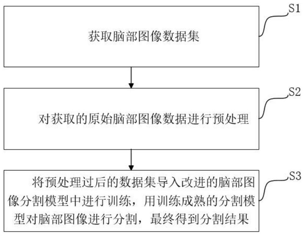
[0030] A brain image segmentation method based on deep learning, comprising:
[0031] S1: Obtain the original brain image dataset;
[0032] Brain MRI image data and segmented hippocampal label image data were obtained from the Alzheimer's Disease Neuroimaging Initiative (ADNI for short) library. The above image data included real patients and healthy comparison groups, and the data format was nifti.
[0033] S2: Preprocessing the acquired original brain image dataset;
[0034] The images in the brain image data set are rotated, mirrored, flipped, and color dithered to enhance processing, cropped, and the image resolution is adjusted, and the brain image data set is divided into a training set and a test set according to the ratio of 8:2.
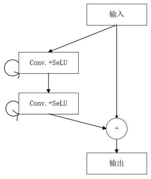
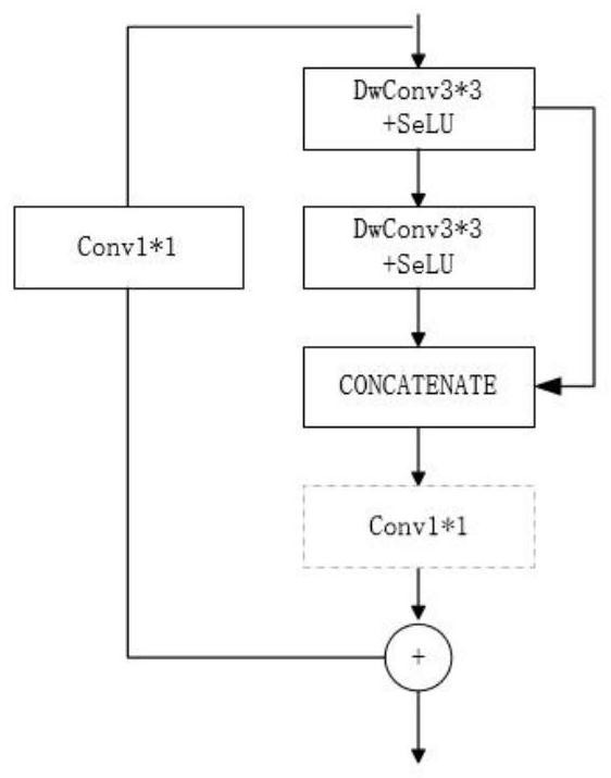
[0035] S3: Import the preprocessed brain image dataset into the brain image segmentation model for training, use the trained and mature brain image segmentation model to segment the brain image, and finally obtain the segmentation result; ...
PUM
 Abstract
Description
Claims
Application Information