An abdominal muscle labeling method and device based on deep learning

A deep learning and muscle technology, applied in the field of medical image processing and artificial intelligence, to achieve the effect of strong practicability, accurate data, and guaranteed accuracy

Active Publication Date: 2019-04-23
ZHONGSHAN HOSPITAL FUDAN UNIV +1
View PDF7 Cites 9 Cited by
  • Summary
  • Abstract
  • Description
  • Claims
  • Application Information

AI Technical Summary

Problems solved by technology

However, there is no relevant report on abdominal muscle labeling based on deep learning

Method used


Image

  • An abdominal muscle labeling method and device based on deep learning
  • An abdominal muscle labeling method and device based on deep learning
  • An abdominal muscle labeling method and device based on deep learning

Examples


Embodiment 1

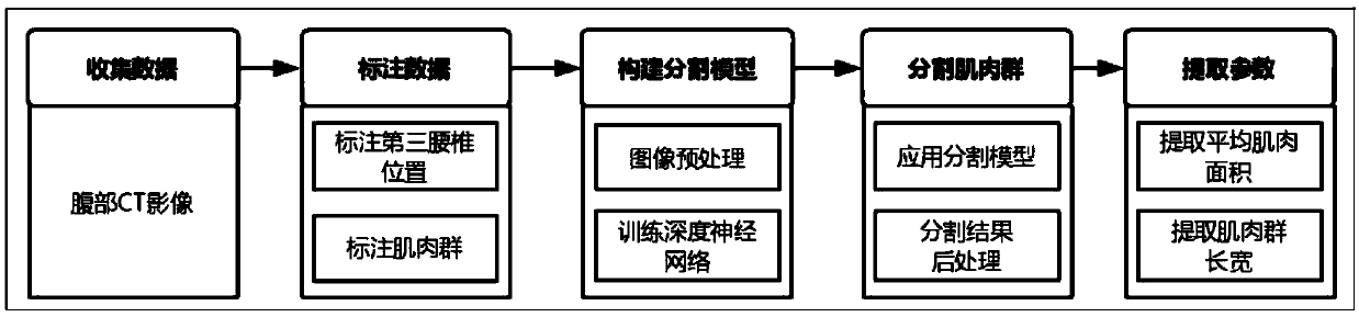
[0057] See figure 1 , figure 1 It is a flowchart of a method for labeling abdominal muscles based on deep learning in the present invention, and the method for labeling abdominal muscles based on deep learning includes the following steps:

[0058] Step 1: Collect Data

[0059] The purpose of this step is to collect abdominal CT image data and guide doctors to collect abdominal CT image data including the third lumbar vertebra. Each CT image contains 5-6 scans of the third lumbar spine.

[0060] In this step, CT data with poor quality (the image of the third lumbar vertebra is too blurred) will be eliminated.

[0061] Step 2: Label the data

[0062] The purpose of this step is to label the abdominal CT image data collected in the first step, and then train the segmentation model. This step includes two sub-steps: marking the position of the third lumbar vertebra and marking muscle groups.

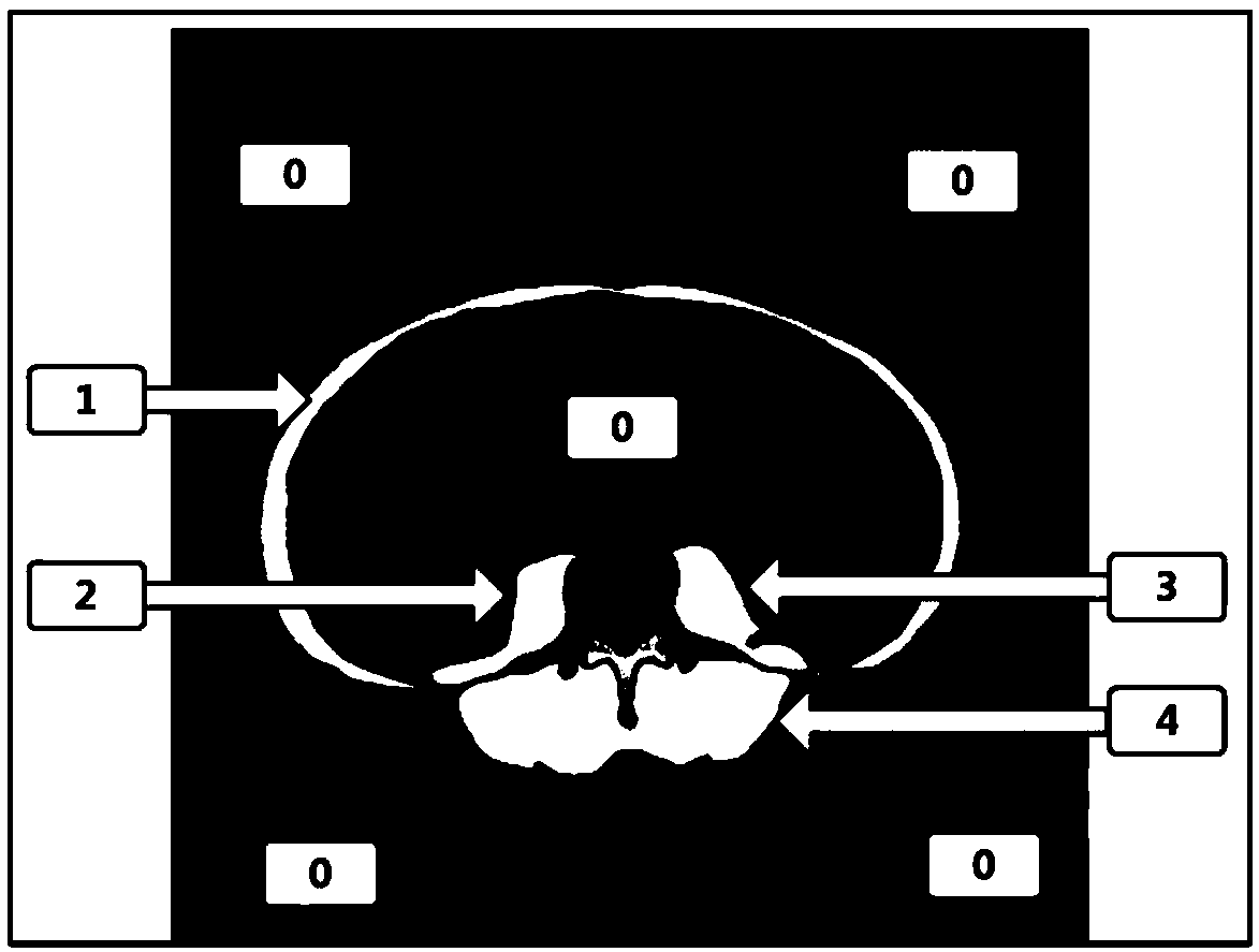
[0063] Mark the position of the third lumbar vertebra, that is, mark the starting...

Embodiment 2

[0079] The abdominal muscle labeling method based on deep learning provided in this embodiment specifically includes the following steps:

[0080] Step 1: Collect Data

[0081] The purpose of this step is to collect abdominal CT image data and guide doctors to collect abdominal CT image data including the third lumbar vertebra. Each CT image contains 5-6 scans of the third lumbar spine.

[0082] In this step, CT data with poor quality (the image of the third lumbar vertebra is too blurred) will be eliminated.

[0083] Step 2: Label the data

[0084] The purpose of this step is to label the abdominal CT image data collected in the first step, and then train the segmentation model. This step includes two sub-steps: marking the position of the third lumbar vertebra and marking muscle groups.

[0085] Mark the position of the third lumbar vertebra, that is, mark the starting CT page number of the third lumbar vertebra. This information will be used in validation testing of th...

Embodiment 3

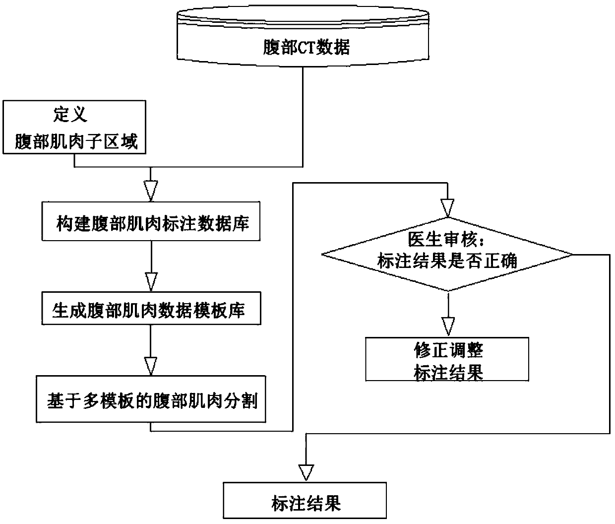
[0109] See Figure 5 , Figure 5 It is a structural block diagram of an abdominal muscle labeling device based on deep learning in the present invention, and the abdominal muscle labeling device based on deep learning includes:

[0110] The image acquisition module 1 is configured to acquire abdominal CT image data including the third lumbar vertebra.

[0111] An annotation module 2 is configured to annotate the abdominal CT image data acquired by the image acquisition module 1 . The marking module 2 further includes two sub-modules, specifically the third lumbar position marking sub-module 21 and the muscle group marking sub-module 22, the third lumbar position marking sub-module 21 is used to mark the third lumbar position, that is, marking the third lumbar position. The starting CT page number of the three lumbar vertebrae. This information will be used in validation testing of the segmentation model. The muscle group labeling sub-module 22 is used for labeling muscle g...


PUM


Abstract

The invention relates to an abdominal muscle labeling method and device based on deep learning. The method comprises the following steps of collecting the abdominal CT image data containing a third lumbar vertebra; marking a third lumbar vertebra position and a muscle group position, wherein four muscle group areas are marked as 1, 2, 3 and 4 respectively, and other areas are marked as 0; generating a label image corresponding to the original CT image, wherein the value of each pixel in the label image is one of {0, 1, 2, 3 and 4}; utilizing the labeled CT image to train a segmentation model,dividing pixels in the CT image into five classes by the segmentation model, and enabling the five classes of pixels to respectively correspond to the labels 0, 1, 2, 3 and 4 in the second step; segmenting the muscle group to obtain the label prediction corresponding to each pixel position in the image; and based on the muscle group segmentation result, calculating the muscle area and the image omics characteristics of the muscle. The device includes the related modules that implement the method. By utilizing the method, the parameters related to the nutrition assessment can be simply, conveniently, quickly and accurately extracted.

Description

technical field [0001] The invention relates to the technical fields of medical image processing and artificial intelligence, in particular to a method and system for marking abdominal muscles based on deep learning. Background technique [0002] The muscle state of the third lumbar vertebrae in the abdomen is an important indicator to measure the nutritional status of a person. Abdominal CT examination is the main means to obtain images of abdominal muscles and then analyze their nutritional status. During the examination, it is necessary to outline the muscle area of ​​the third lumbar vertebrae in the CT image, and then calculate the relevant parameters to judge the nutritional status. The current conventional method is that the doctor manually outlines the area, and then calculates relevant parameters (commonly used parameters include muscle area, length and width, etc.). Although conventional methods can obtain accurate muscle regions and parameters, they bring a great...

Claims


Application Information

Patent Timeline
Patent Type & Authority Applications(China)
IPC IPC(8): G06T7/00G06T7/174
CPCG06T7/0012G06T7/174G06T2207/10081G06T2207/20084G06T2207/20081
Inventor 刘迎迎陈世耀周继
Owner ZHONGSHAN HOSPITAL FUDAN UNIV
Why Eureka
Try Eureka