A kind of pathological image classification method and computer equipment

A technology of pathological images and classification methods, applied in image analysis, computer components, computer-aided medical procedures, etc. Fitting and other issues to achieve the effect of improving classification accuracy, realizing classification tasks, and preventing overfitting

Active Publication Date: 2020-12-11
SHENZHEN UNIV
View PDF6 Cites 0 Cited by
  • Summary
  • Abstract
  • Description
  • Claims
  • Application Information

AI Technical Summary

Problems solved by technology

The inventors found that the above-mentioned methods only use a single deep convolutional neural network (DCNN), which can neither obtain the shallow features (texture, color, shape, etc.) and deep semantic features of the image at the same time, nor use feature selection algorithms. Removing noise and redundant information is prone to overfitting problems, which seriously affects the accuracy and robustness of the system

Method used


Image

  • A kind of pathological image classification method and computer equipment
  • A kind of pathological image classification method and computer equipment
  • A kind of pathological image classification method and computer equipment

Examples


Embodiment 1

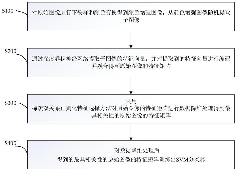
[0045] figure 1 A schematic flowchart of the pathological image classification method provided by the first embodiment of the present invention is shown.

[0046] Step S100, performing down-sampling and color transformation on the original image to obtain a first predetermined number of color-enhanced images, and randomly extracting a second predetermined number of sub-images from each of the color-enhanced images;

[0047] As mentioned above, the pathological image is a pathological image of breast cancer;

[0048] Optionally, the size of the original image of breast cancer is 2048×1536 pixels, and the downsampling is performed to reduce its size to half of the original image (1024×768 pixels) as the input image, which avoids the excessive size of the image and leads to pre-training The incompatibility of the network, and allowing the selection of smaller sub-image sizes without causing loss of important feature information; then transforming the original image from RGB spac...

Embodiment 2

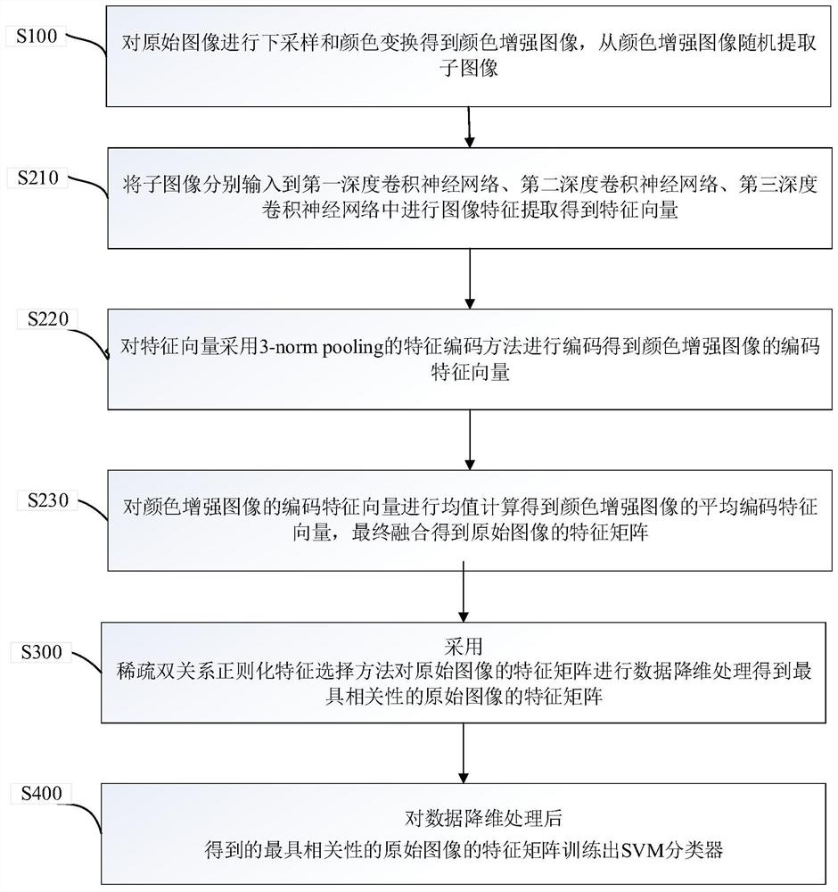
[0059] refer to figure 2 , the second embodiment of the present invention provides a pathological image classification method, based on the above figure 1 In the first embodiment shown, the step S200 "extracts the feature vectors of the sub-images respectively through a third predetermined number of deep convolutional neural networks, and extracts the feature vectors of the third predetermined number of deep convolutional neural networks to The eigenvectors of the original image are encoded and fused to obtain the eigenmatrix of the original image, including:

[0060] Step S210, respectively input the sub-images into the first deep convolutional neural network, the second deep convolutional neural network, and the third deep convolutional neural network for image feature extraction to obtain the feature vector;

[0061] As mentioned above, the third predetermined number is three, which are the first deep convolutional neural network, the second deep convolutional neural netw...

Embodiment 3

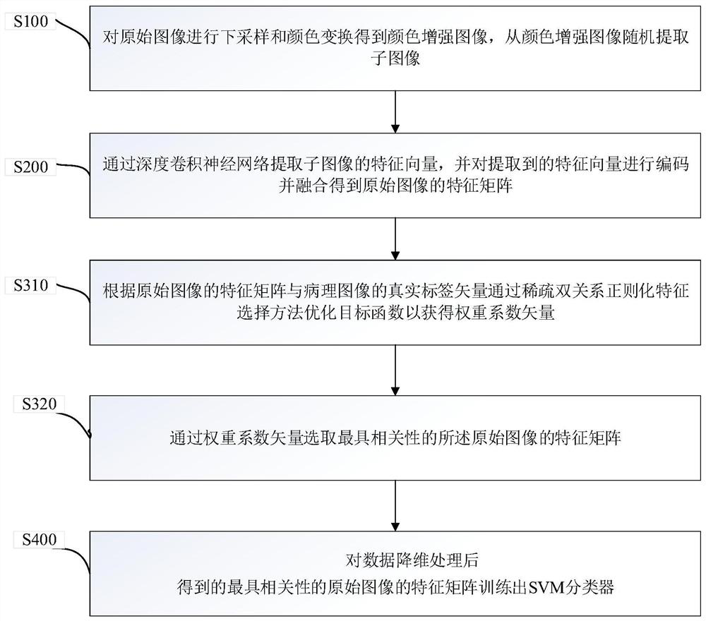
[0078] refer to image 3 , the third embodiment of the present invention provides a pathological image classification method, based on the above figure 1 In the first embodiment shown, the step S300 of "using the sparse dual-relational regularization feature selection method to perform data dimensionality reduction processing on the feature matrix of the original image to obtain the most relevant feature matrix of the original image" includes:

[0079] S310, according to the feature matrix of the original image and the real label vector of the pathological image, the objective function is optimized by a sparse double-relational regularization feature selection method to obtain a weight coefficient vector, wherein the objective function is expressed as:

[0080]

[0081] where w is the weight coefficient vector; y is the real label vector of the original image;

[0082] Specifically, the objective function optimized by the sparse dual-relational regularization feature selec...


PUM


Abstract

The invention discloses a pathological image classification method and computer equipment. According to the method, performing down-sampling and color transformation on an original image to obtain anenhanced image, randomly extracting sub-images from the color enhanced image, and reserving original image information as much as possible; extracting feature vectors of the sub-images through a deepconvolutional neural network, wherein the feature vectors comprise deep semantic features of the images and textures, colors, shapes and other features of shallow layers; encoding and fusing the feature vectors to obtain a feature matrix of the original image; and carrying out data dimension reduction processing on the feature matrix of the original image by adopting a sparse double-relation regularization feature selection method to obtain a feature matrix of the original image with the highest correlation, and training an SVM classifier according to the feature matrix of the original image with the highest correlation to realize a classification task of the pathological image. By means of the method, comprehensive original image feature information can be obtained, and an accurate classification task can be achieved under limited training data.

Description

technical field [0001] The present invention relates to the technical field of medical image classification, in particular to a pathological image classification method and computer equipment. Background technique [0002] Breast cancer is one of the most important malignant tumors affecting women's health. The incidence rate varies significantly across regions around the world. The incidence of breast cancer in developed countries and regions is significantly higher than that in less developed countries and regions, ranking first in the death of female malignant tumors. At the same time, the number of cases and deaths of breast cancer in Chinese women accounted for 11.2% and 9.2% of the global incidence and death respectively, ranking among the top in the world. Therefore, how to improve the accuracy of early diagnosis of breast cancer and reduce the mortality rate of cancer is very important for the development of medicine. At present, early diagnosis of breast cancer is...

Claims


Application Information

Patent Timeline
Patent Type & Authority Patents(China)
IPC IPC(8): G06K9/62G06K9/66G06N3/04G06T5/00G06T7/00G06T7/90G16H50/20
Inventor 汪天富王永军雷柏英陈思平
Owner SHENZHEN UNIV
Why Eureka
Try Eureka