
Organ volume detection method and device

A detection method and organ technology, applied in instruments, image analysis, image enhancement, etc., can solve the problems of inaccurate organ volume detection and large image error, etc.

Active Publication Date: 2021-04-20
SHANGHAI CHANGZHENG HOSPITAL
View PDF15 Cites 0 Cited by
  • Summary
  • Abstract
  • Description
  • Claims
  • Application Information

AI Technical Summary

Problems solved by technology

[0005] The object of the present invention is to provide a method and device for organ volume detection in view of the deficiencies in the prior art above, so as to solve the problem of inaccurate organ volume detection due to large image errors of organs acquired in the related art The problem

Method used

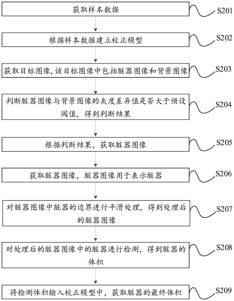
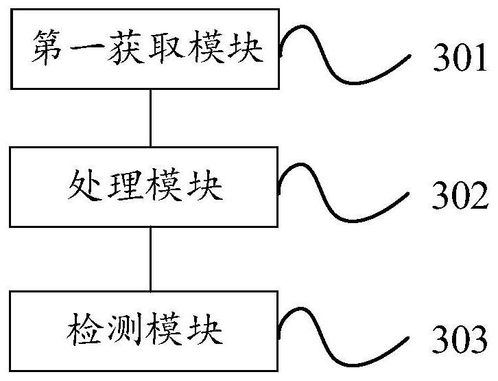

Image

  • Organ volume detection method and device
  • Organ volume detection method and device
  • Organ volume detection method and device

Examples


Embodiment Construction

[0055] In order to make the purpose, technical solutions and advantages of the embodiments of the present invention clearer, the technical solutions in the embodiments of the present invention will be clearly and completely described below in conjunction with the drawings in the embodiments of the present invention. Obviously, the described embodiments It is a part of embodiments of the present invention, but not all embodiments.

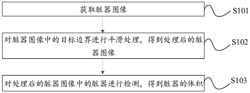
[0056] figure 1 A schematic flow chart of the organ volume detection method provided by an embodiment of the present invention, such as figure 1 As shown, the method includes:

[0057] Step 101, acquiring organ images.

[0058] In order to prevent some diseases caused by the change of organ volume, it is necessary to check the patient's organs to obtain the target image including the organ image. After the target image is segmented, the organ image can be obtained, so that the obtained organ The image of the organ can be used to judge whether the...


PUM


Abstract

The invention provides an organ volume detection method and device, which relate to the technical field of medical organ detection. Including: obtaining the organ image, which is used to represent the organ; smoothing the target boundary in the organ image to obtain the processed organ image, the target boundary is the organ obtained through the segmentation operation triggered by the user The boundary of the organ; finally, detect the organ in the processed organ image to obtain the volume of the organ. In the embodiment of the present invention, the error of the acquired image of the organ is reduced by smoothing the target boundary in the organ image, so that the volume detection of the organ is more accurate.

Description

technical field [0001] The present invention relates to the technical field of medical organ detection, in particular to an organ volume detection method and device. Background technique [0002] In medicine, organs refer to internal organs such as the heart, intestines, lungs, liver, spleen, and kidneys. Through the calculation of organ volume, it can be used to prevent the occurrence of some diseases, and at the same time, it also plays a vital role in the diagnosis and treatment of some diseases. [0003] In the related technology, the medical personnel obtain the nuclear magnetic resonance image containing the shape of the organ through the nuclear magnetic resonance technology, and then segment the image through the terminal to obtain the image of the organ, and the terminal can correspond to the image according to the image of the organ. The volume of the organ is calculated to obtain the volume of the organ corresponding to the image. [0004] However, when an image...

Claims


Application Information

Patent Timeline
Patent Type & Authority Patents(China)
IPC IPC(8): G06T7/62G06T5/00G06T7/12G06T7/136
Inventor 宋书伟梅长林付莉莉苏蓓琳吕佳颐
Owner SHANGHAI CHANGZHENG HOSPITAL
Learn More