Minimally invasive visual expansion attraction sheath

A technique of attracting sheath and dilating tube, applied in the direction of puncture needle, trocar, etc., can solve problems such as renal bleeding, and achieve the effect of eliminating renal bleeding and bladder bleeding

Pending Publication Date: 2019-06-28
海盐康源医疗器械有限公司
View PDF0 Cites 2 Cited by
  • Summary
  • Abstract
  • Description
  • Claims
  • Application Information

AI Technical Summary

Problems solved by technology

[0003] However, the traditional percutaneous nephroscope/flexible ureteroscope also has defects. For example, when dilation is performed after the puncture, the traditiona...

Method used


Image

  • Minimally invasive visual expansion attraction sheath
  • Minimally invasive visual expansion attraction sheath
  • Minimally invasive visual expansion attraction sheath

Examples


Embodiment Construction

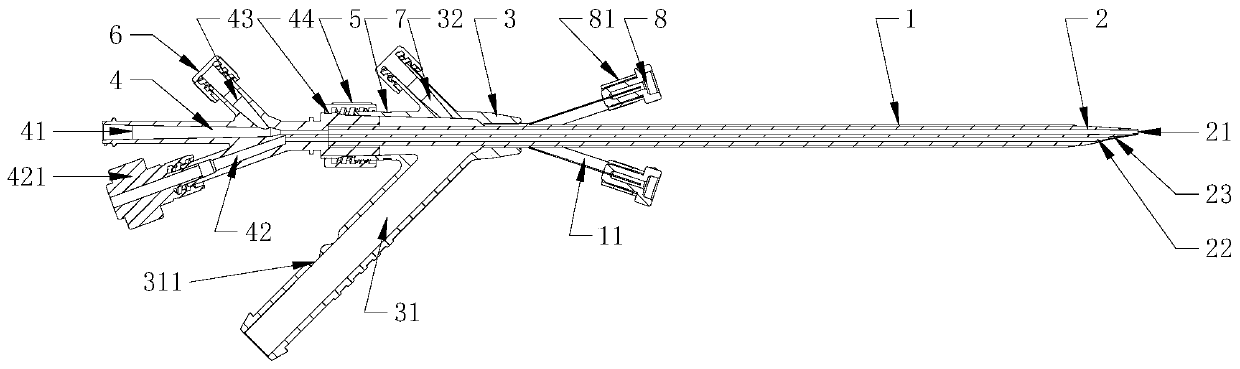
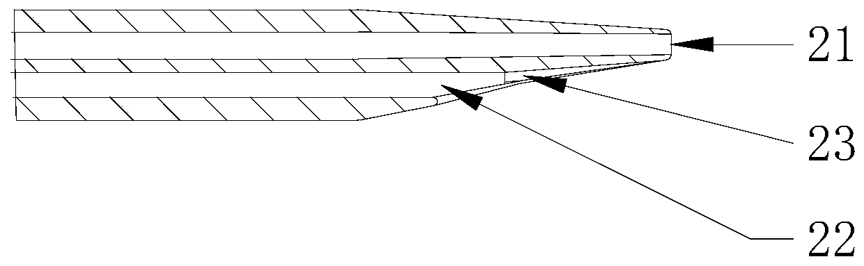
[0024] refer to Figure 1 to Figure 5 , a minimally invasive visual dilation suction sheath of the present invention, comprising a sheath tube 1, a dilation tube 2, a sheath tube joint 3, a dilation tube joint 4 and a tightening joint 5, the dilation tube 2 is located inside the sheath tube 1 and is connected to the sheath tube 1 The sheath tube 1 is coaxial, and the sheath tube joint 3 and the expansion tube joint 4 are connected and fixed by screwing the joint 5, and one end of the sheath tube 1 is fixed on the main channel of the sheath tube joint 3, and the expansion tube 2 One end is in the shape of a truncated cone and the other end is fixed on the main channel of the expansion tube joint 4. The first optical fiber channel 21 and the first guide wire channel 22 in the expansion tube 2 are connected with the second optical fiber channel 41 in the expansion tube joint 4 respectively. It communicates with the second guide wire channel 42, and the expansion tube joint 4 is p...


PUM


Abstract

The invention discloses a minimally invasive visual expansion attraction sheath. The minimally invasive visual expansion attraction sheath includes a sheath tube, an expansion tube, a sheath tube connector, an expansion tube connector and a tightening connector. The expansion tube is located in the sheath tube and coaxial with the sheath tube, the sheath tube is fixedly connected with the expansion tube connector through the tightening connector, one end of the sheath tube is fixed on a main channel of the sheath tube connector, one end of the expansion tube is in a circular truncated cone shape, the other end of the expansion tube is fixed on a main channel of the expansion tube connector, and a first optical fiber channel and a first wire guide channel in the expansion tube are communicated with a second optical fiber channel and a second wire guide channel in the expansion tube connector. At the time of expansion and puncture, a visual image system can be applied to effectively locate puncture and expansion positions, and medical accidents such as renal hemorrhage and bladder hemorrhage caused by deep puncture in a routine operation are prevented.

Description

【Technical field】 [0001] The invention relates to the technical field of medical devices, in particular to the technical field of a minimally invasive visible expansion suction sheath. 【Background technique】 [0002] Traditional treatment methods for kidney stones include incision lithotripsy, extracorporeal lithotripsy, and percutaneous nephrolithotomy. Traditional treatment methods for bladder stones include extracorporeal shock wave lithotripsy and suprapubic bladder surgery. The open "surgery to remove stones" method is to make a large incision at the stone site, and then take out the stones, which has the disadvantages of large damage, more bleeding, heavy pain, and slow recovery. After the operation, the patient usually needs to be hospitalized for about 7 days, because the surgical incision is large, not only the skin is not beautiful, but the physical strength of the patient is greatly affected after the operation. Compared with open surgery, percutaneous nephroscop...

Claims


Application Information

Patent Timeline
IPC IPC(8): A61B17/34
Inventor 程跃郁鹏忠钱江史军周伟
Owner 海盐康源医疗器械有限公司
Why Eureka
Try Eureka