Deep learning method for prostate cancer auxiliary diagnosis

A technology for auxiliary diagnosis and prostate cancer, applied in the field of deep learning, can solve the problems of low precision and high time-consuming of prostate tissue segmentation, achieve the effect of improving segmentation effect, accurate segmentation result, and improving segmentation efficiency

Active Publication Date: 2019-07-12
HEFEI UNIV OF TECH
View PDF4 Cites 5 Cited by
  • Summary
  • Abstract
  • Description
  • Claims
  • Application Information

AI Technical Summary

Problems solved by technology

[0004] The purpose of the present invention is to provide a deep learning method for the auxiliary diagnosis of prostate cancer to solve the problems of low accuracy and high time-consuming for prostate tissue segmentation in the prior art

Method used


Image

  • Deep learning method for prostate cancer auxiliary diagnosis
  • Deep learning method for prostate cancer auxiliary diagnosis
  • Deep learning method for prostate cancer auxiliary diagnosis

Examples


Embodiment Construction

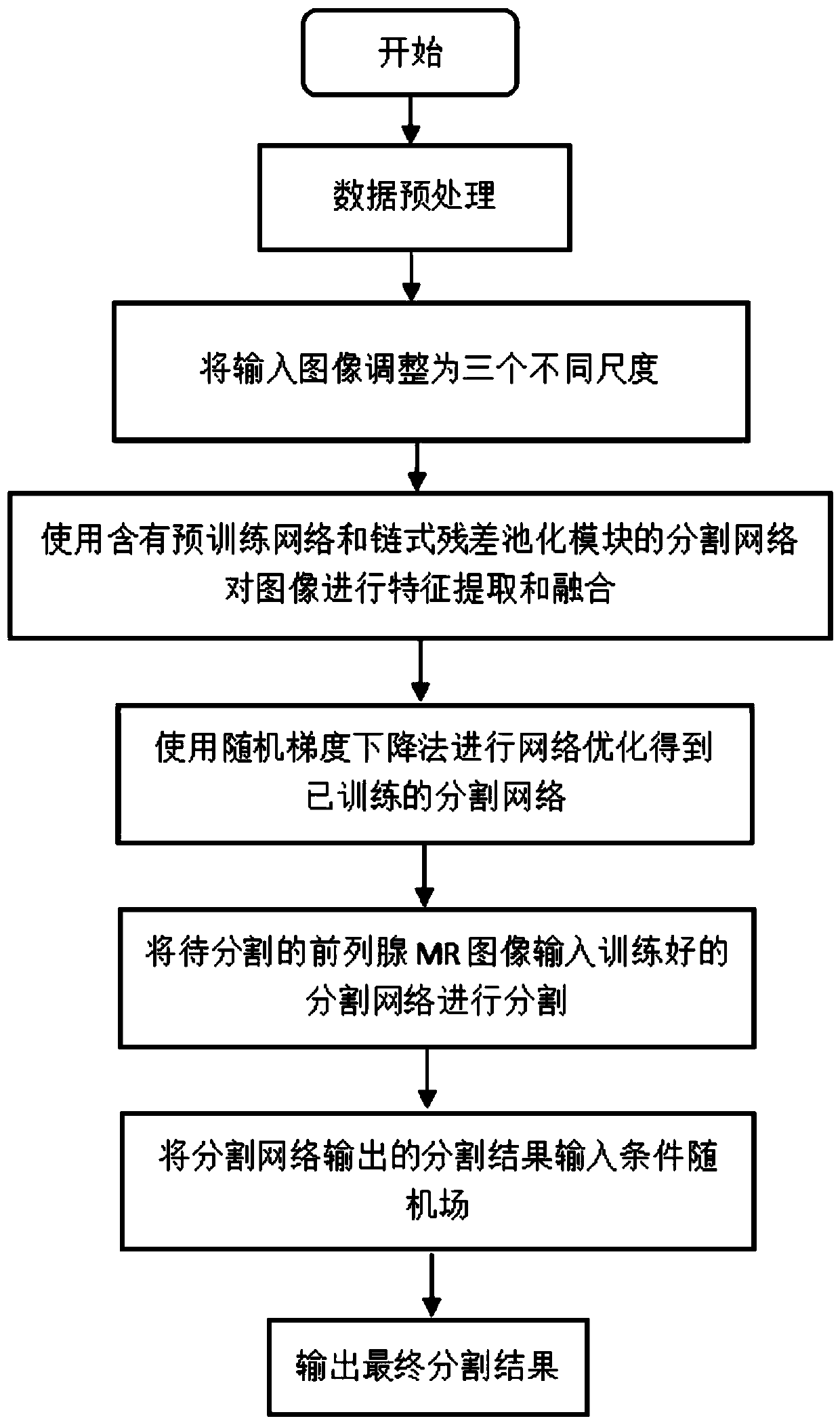
[0023] Such as figure 1 As shown, a deep learning method for auxiliary diagnosis of prostate cancer, the process is as follows:

[0024] (1), select 686 pieces of prostate MR images of 45 patients and the artificial segmentation map of corresponding prostate tissue as the training data set;

[0025] (2) Preprocess the data set, expand the data set by horizontal and vertical flipping and adjust brightness, contrast, and saturation data enhancement methods, and expand the training pictures according to the original picture {1, 0.75, 0.5 respectively } is resized to 3 scales;

[0026] (3) Input the multi-scale image obtained in step (2) into the segmentation network model for training. The segmentation network is mainly composed of a ResNet pre-training model and a chained residual pooling module. The pictures of the three scales are respectively input into a ResNet pre-training model, and the multi-scale features of the input image are extracted by fine-tuning the parameters of...


PUM


Abstract

The invention discloses a deep learning method for prostate cancer auxiliary diagnosis. The method comprises: establishing a segmentation network model containing a pre-training model and a chain typeresidual pooling module; carrying out feature extraction and fusion on the input prostate MR images with different scales; and then optimizing the constructed segmentation network by using a random gradient descent algorithm to obtain a trained model, inputting the prostate MR image to be segmented into the trained model, and finally outputting a final segmentation result from a result output bythe model through a conditional random field. The segmentation result of the method can assist doctors in clinical diagnosis and treatment of the prostatic cancer, and the diagnosis accuracy and the working efficiency of the doctors are effectively improved.

Description

technical field [0001] The invention relates to the field of deep learning methods, in particular to a deep learning method for auxiliary diagnosis of prostate cancer. Background technique [0002] Today, prostate cancer has become a major threat to the health of men, especially older men, and is one of the most common cancers in recent decades. When clinically diagnosing prostate cancer, doctors need to separate the prostate tissue from the surrounding tissues and organs in prostate MR images for easy diagnosis and treatment. The meaningful information extracted by this segmentation process includes shape, relative position of organs, volume and abnormalities. Although the contrast between soft tissue organs in MR images is better than that in computed tomography, less valid information is available due to the small area belonging to prostate tissue in MR images, and the size, shape, and location of prostate tissue in each patient are different. vary, and accurate delinea...

Claims


Application Information

Patent Timeline
Patent Type & Authority Applications(China)
IPC IPC(8): G06K9/62G16H30/00
CPCG16H30/00G06F18/253G06F18/214Y02T10/40
Inventor 詹曙陈爱莲臧怀娟
Owner HEFEI UNIV OF TECH
Why Eureka
Try Eureka