Micro-immunofluorescence detection method without cell loss

An immunofluorescence detection and cell-free technology, which is applied in the biological field, can solve the problems of limiting the application of immunofluorescence technology and difficult identification, and achieve the effects of improving visibility, reducing experimental background, and eliminating cell loss

Active Publication Date: 2019-08-06
ZHEJIANG UNIV
View PDF12 Cites 4 Cited by
  • Summary
  • Abstract
  • Description
  • Claims
  • Application Information

AI Technical Summary

Benefits of technology

This patented method uses specialized reagents called primers specifically target cancerous tissue cells from healthy individuals' circulating lymphatic system. These primer molecules are attached to various substances like albumen, collagen, gelatin, etc., making them easy targets for imagining these areas during histopathology analysis. By selecting appropriate ones for each patient by measuring their own unique gene expression levels, this technique allows researches looking at how well they work out over time without sacrificing accuracy. Overall, this process provides technical means for identifying and analyzing rare diseases such as autoimmune disorders through flow cytometry techniques.

Problems solved by technology

Technologies have developed overall, these technics aim to provide better ways to detect diseases like cancer through various means including immune fibroupletance assay(IFA). These include immunofungluximab (FIS)-based tests, flowcytelectron emission tomography (FECT), quantum dots, and laser scanning imagers. While IFAs allow precise detection of disease states within living organisms, their limitations exist due to factors like weakness caused by natural fixations between different parts of organoids called chromatophoric receptors. Fluorescences offer improved accuracy compared to conventional FIBS while being more sensitive than optically based systems. Additionally, there may still remain issues associated with current immunosuppressive procedures involving enzyme activation and deposition.

Method used


Image

  • Micro-immunofluorescence detection method without cell loss
  • Micro-immunofluorescence detection method without cell loss
  • Micro-immunofluorescence detection method without cell loss

Examples


Embodiment 1

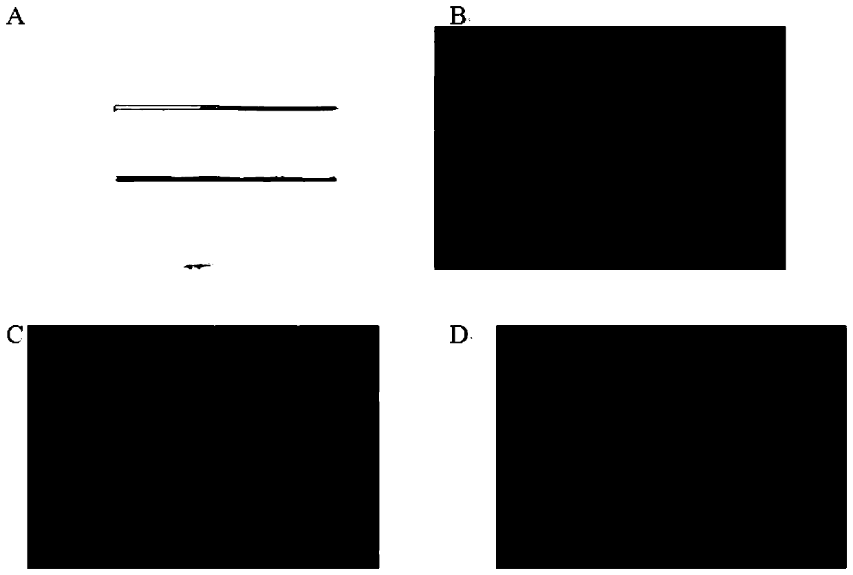
[0106] Embodiment 1. A micro-immunofluorescence detection method without cell loss, the following steps are carried out in sequence:

[0107] 1) Sterilize the 0.22um Spin-X Centrifuge Tube with high-pressure steam (pressure of 103.4KPa, temperature of 121°C, sterilization time of 30 minutes);

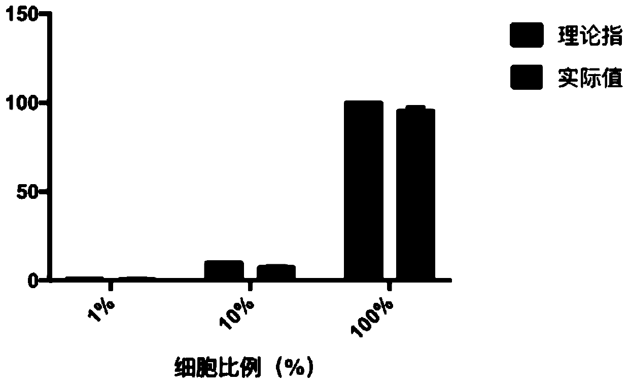
[0108] 2), using a cell counting board to take a certain number of A549 cells and 3T3 cells to mix, respectively to obtain the following experimental groups:

[0109] Experimental group 1: the ratio of A549 cells is 1% (ie, A549 cells: 3T3 cells = 1:99);

[0110] Experimental group 2: the ratio of A549 cells is 10% (ie, A549 cells: 3T3 cells = 1:9);

[0111] Experimental group 3: the proportion of A549 cells is 100% (that is, all are A549 cells);

[0112] The above three experimental groups all meet the following conditions: the total number of cells is 1*10 5 pcs; the volume is 0.5ml;

[0113] The above-mentioned 3 experimental groups were respectively carried out as follows: trans...

Embodiment 2

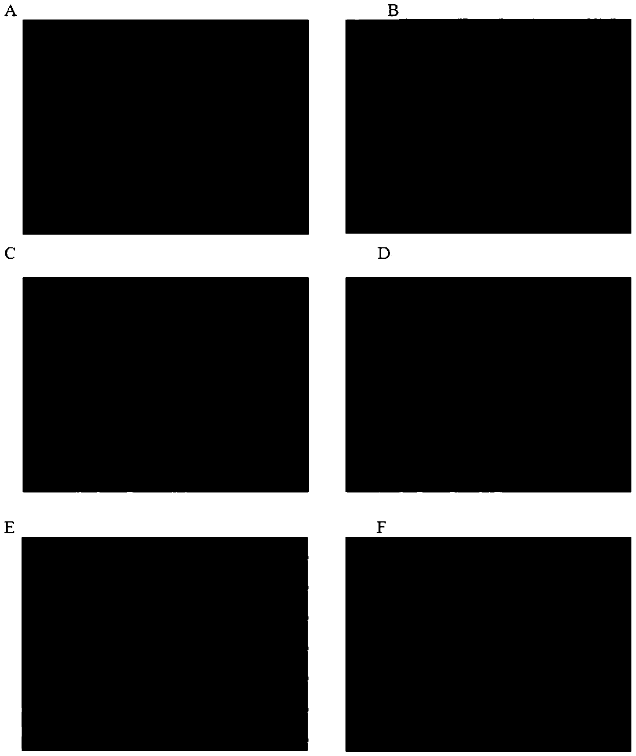
[0144] Embodiment 2: Compared with Embodiment 1, only step 2 is changed; the rest is the same as Embodiment 1.

[0145] 2) Set up the following experimental groups respectively:

[0146] Experimental group 1 (negative control experiment): 0.5ml of cell culture medium was directly transferred to the sterilized 0.22um Spin-X Centrifuge Tube obtained in step 1);

[0147] Experimental group 2: the adherent cells --- mouse fibroblast 3T3 cells (concentration: 1*10 4 ) 0.5ml is directly transferred to the sterilized 0.22um Spin-X Centrifuge Tube obtained in step 1);

[0148] Experimental group 3: non-adherent cells - human acute leukemia cells HL60 (concentration 1*10 4 ) 0.5ml is directly transferred to the sterilized 0.22um Spin-X Centrifuge Tube obtained in step 1);

[0149] Experimental group 4: non-adherent cells - human acute leukemia cells HL60 (concentration 1*10 4) 0.5ml is transferred to the sterilized 0.22um Spin-X Centrifuge Tube obtained in step 1) and cultivated in...


PUM


Abstract

The invention discloses a micro-immunofluorescence detection method without cell loss. The micro-immunofluorescence detection method comprises the steps of: performing 0.22 um Spin-X Centrifuge Tube sterilization; transferring cells into a sterilized 0.22 um Spin-X Centrifuge Tube for carrying out subsequent steps directly or carrying out subsequent steps fter cultivation; and performing the subsequent steps of PBS buffer solution rinsing, 4% paraformaldehyde fixation, methanol treatment on ice, 0.3 M glycine confining liquid treatment, 4% BSA confining liquid treatment, fluorescence primary antibody room temperature dark incubation, staining in DAPI staining solution, slide sealing after placing a filtering membrane in a slide, and observation of the slide under a laser scanning cofocal microscope. The method makes up for the shortcoming that the immunofluorescence technique is difficult to achieve precise screening of specific cells, and provides a simple, precise and visual technique for screening specific cells in a mixed cell population.

Description


Claims


Application Information

Patent Timeline
Owner ZHEJIANG UNIV
Why Eureka
Try Eureka