Clinical fixation and operating device of B-mode ultrasound probe for B-mode ultrasound room

A manipulation device, clinical technology, applied in applications, ultrasonic/sonic/infrasonic diagnosis, medical science, etc., can solve the problems of missing fine pictures, affecting the diagnosis results, unable to make large-scale adjustments, etc., to achieve position adjustment Accurate and stable, fast installation and split work, quick positioning and effect of scanning work

Inactive Publication Date: 2019-08-16
THE AFFILIATED HOSPITAL OF QINGDAO UNIV
View PDF1 Cites 1 Cited by
  • Summary
  • Abstract
  • Description
  • Claims
  • Application Information

AI Technical Summary

Problems solved by technology

[0002] At present, when a patient needs to undergo B-ultrasound examination, the patient lies on the examination bed, and the B-ultrasound doctor needs to hold the B-ultrasound probe in his hand and move it continuously in the patient's abdomen to check the patient's body parts, and then observe the B-ultrasound display device. Image, the previous method has the following disadvantages when the patient undergoes B-ultrasound examination: first, the B-ultrasound doctor keeps looking for the image while holding the B-ultrasound probe, and observes the displayed image at the same time, which is time-consuming and laborious; second, the B-ultrasound doctor The B-ultrasound probe cannot be stabilized very well, so that the picture is not clear and affects the diagnosis result; third, the B-ultrasound doctor keeps moving the B-ultrasound probe and observing the image during the examination, which is easy to be distracted and miss the fine picture, increasing the Increased the difficulty of the work of B-ultrasound physicians
[0004] But a kind of B-ultrasound room clinical use B-ultrasound probe fixed manipulation device provided in the above-mentioned technical scheme still has many disadvantages in actual use. Scanning detection is performed, and the height and weight of the inspector will also affect the detection requirements, and large-scale adjustments cannot be made

Method used


Image

  • Clinical fixation and operating device of B-mode ultrasound probe for B-mode ultrasound room
  • Clinical fixation and operating device of B-mode ultrasound probe for B-mode ultrasound room
  • Clinical fixation and operating device of B-mode ultrasound probe for B-mode ultrasound room

Examples


Embodiment approach

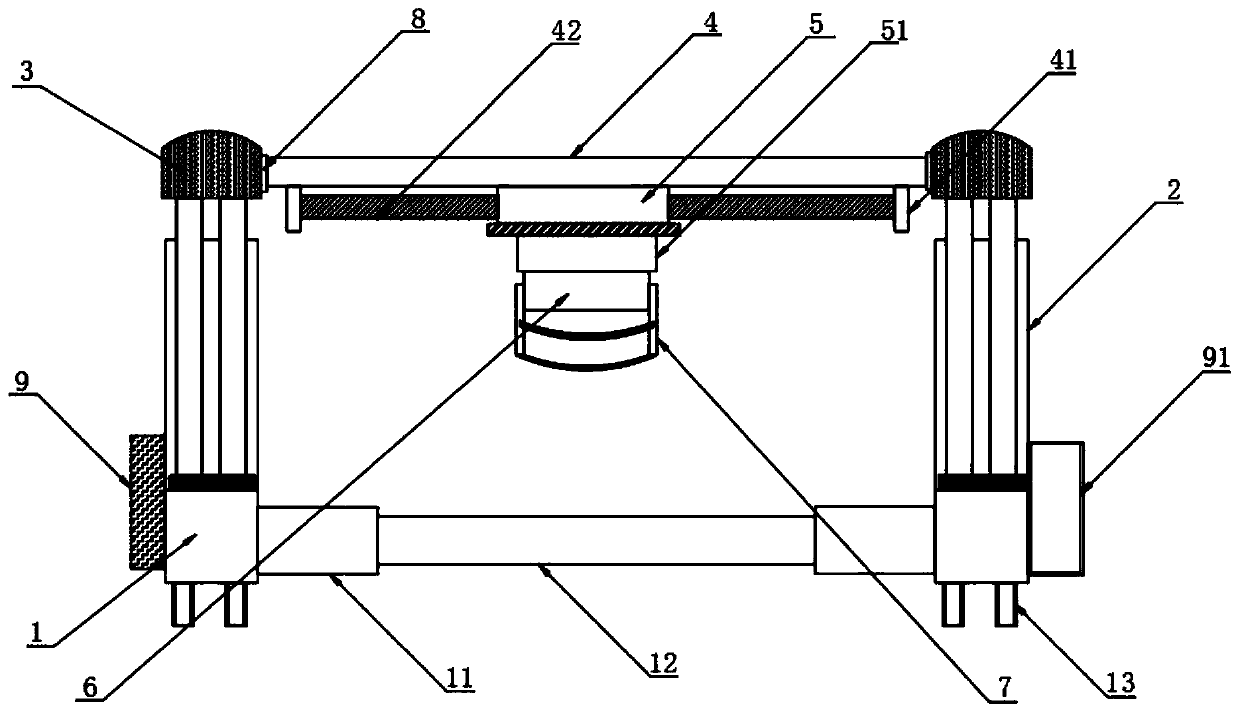
[0037] The implementation method is as follows: put the B-ultrasound probe inside the positioning seat 6, and then use the fastening belt 72 connected to the outside of the positioning plate 7 to connect the positioning plate 7 and the B-ultrasound probe tightly, so as to realize simple and fast positioning work, and the positioning is completed After controlling the operation of the B-ultrasound probe and the driving motor, the rapid positioning and scanning of the B-ultrasound probe can be realized.

[0038] The inside of the data transmission device 9 is provided with a PLC controller, a wireless data transmission module and a data processing module, the outside of the data transmission device 9 is connected with a data transmission cable, and the other side surface of the base 1 is provided with a storage box 91, The data transmission device 9 is connected to the computer on which the medical personnel view the data through a transmission cable, and can receive control sign...


PUM


Abstract

The invention discloses a clinical fixation and operating device of a B-mode ultrasound probe for a B-mode ultrasound room and particularly relates to the field of medical assist devices. The clinicalfixation and operating device comprises two bases; a pillar is arrange at the top of each base; a top seat is arranged at the top of each pillar; an ejector bar is arranged between the two bases; a sliding seat is arranged at the bottom of the ejector bar; a motor is arranged at the bottom of the sliding seat; a positioning seat is connected to the outer side of the motor and is in drive connection with the motor. Moving seats, the pillars and a driving mechanism are provided; adjusting is performed during use in accordance with different parts which an B-mode ultrasound probe is to scan; theheight of the B-mode ultrasound probe can be adjusted by controlling first hydraulic lifting bars to operate; the transverse position of the B-mode ultrasound probe can be adjusted by controlling anelectric push bar to operate; the vertical position of the B-mode ultrasound probe can be adjusted by controlling the driving motor to operate; adjusting is performed simply and quickly; medical personnel can check out B-mode ultrasound scanning results and data directly on a computer.

Description

technical field [0001] The invention relates to the technical field of medical auxiliary devices, more specifically, the invention relates to a B-ultrasound probe fixing and manipulating device for clinical use in a B-ultrasound room. Background technique [0002] At present, when a patient needs to undergo B-ultrasound examination, the patient lies on the examination bed, and the B-ultrasound doctor needs to hold the B-ultrasound probe in his hand and move it continuously in the patient's abdomen to check the patient's body parts, and then observe the B-ultrasound display device. Image, the previous method has the following disadvantages when the patient undergoes B-ultrasound examination: first, the B-ultrasound doctor keeps looking for the image while holding the B-ultrasound probe, and observes the displayed image at the same time, which is time-consuming and laborious; second, the B-ultrasound doctor The B-ultrasound probe cannot be stabilized very well, so that the pic...

Claims


Application Information

Patent Timeline
IPC IPC(8): A61B8/00
CPCA61B8/4209
Inventor 何香芹
Owner THE AFFILIATED HOSPITAL OF QINGDAO UNIV
Why Eureka
Try Eureka