

Method for producing cartilage cells induced to be differentiated from stem cells
A stem cell differentiation and chondrocyte technology, applied in the field of preparation of chondrocytes induced from stem cell differentiation, can solve problems such as high cost and achieve the effect of high expression level
- Summary
- Abstract
- Description
- Claims
- Application Information
AI Technical Summary
Problems solved by technology
Method used
Image

Examples
Embodiment approach
[0155] Hereinafter, the present invention will be described in detail.
[0156] In the field of regenerative medicine, the technology of inducing the differentiation of chondrocytes by cell therapy technology is widely used for cartilage regeneration, but the therapeutic effect may vary depending on the characteristics of the differentiation ability and tissue formation ability of the transplanted chondrocytes. For this reason, a differentiation technique for inducing chondrocytes derived from reverse-differentiated stem cells is required, and studies are continuing for the purpose of improving differentiation efficiency and tissue-forming ability of differentiated chondrocytes.
[0157] Therefore, the present invention provides a method for preparing chondrocytes induced by stem cell differentiation through the following steps i) to step v):
[0158] Step i), forming (generation) embryoid bodies by culturing reverse differentiated stem cells (induced pluripotent stem cells, i...
Embodiment 1
[0244] Example 1. Generation of human induced pluripotent stem cells using isolated CBMCs
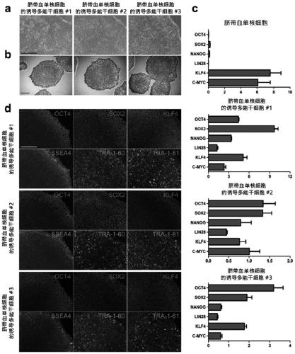
[0245] Reprogramming of CBMCs was facilitated using Sendai virus containing Yamanaka factors. Yamanaka factors are genes that induce pluripotency. After a period of transduction, CBMC-hiPSCs formed colonies similar to embryoid body stem cells ( figure 1 part (a) of this document). CBMC-hiPSCs were purified using a cell line with the same cell morphology. The same CBMC-hiPSCs were used for additional characterization. The identified CBMC-hiPSCs were stained with alkaline phosphate ( figure 1 part (b) of Multidifferentiation markers including OCT4, SOX2, NANOG, LIN28, KLF4, and c-MYC were also assayed ( figure 1 part (c) of Maternal CBMCs were used as a negative control group. The expressions of OCT4, SOX2, NANOG, and LIN28 were increased in CBMC-hiPSCs. However, KLF4 and c-MYC were less expressed in CBMC-hiPSCs than in CBMCs. Typical cell surface markers (SSEA4, OCT4, SOX2, KLF...
Embodiment 2
[0246] Example 2. Chondrocytes differentiated into CBMC-iPSCs
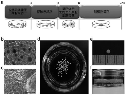
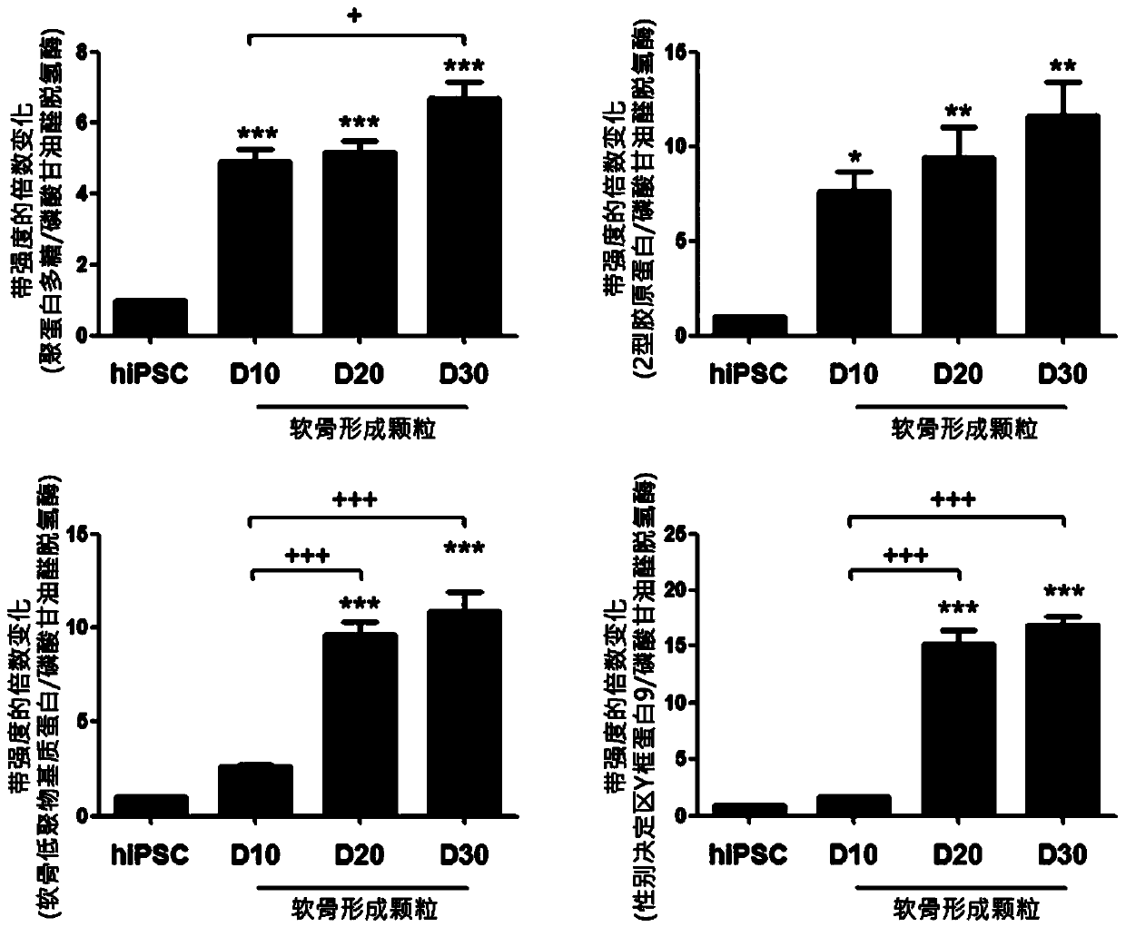
[0247] To confirm the cartilage regeneration ability of CBMC-iPSCs, chondrogenic differentiation was performed by culturing embryoid bodies and inducing growth cells. A simple scheme for the generation process of chondrogenically differentiated cell bodies is shown in figure 2 as shown in part (a). CBMC-iPSCs colonies were prepared for chondrogenic differentiation ( figure 2 part (b) of CBMC-iPSCs expanded and aggregated into embryoid bodies ( figure 2 part (c) of After several days of expansion, the embryoid bodies were transferred to gelatin-coated dishes to induce growth cells ( figure 2 part (d) of this document). Growing cells are expanded and isolated into single cells for chondrocyte differentiation. Utilize 2×10 6iPScs were obtained from many chondrocyte bodies. After 30 days of differentiation, embryoid body growth cells were used to form chondrocyte bodies. The resulting chondrogenic partic...
PUM
 Abstract
Description
Claims
Application Information