Method and apparatus for processing histological image captured by medical imaging device

A medical imaging and histological technology, applied in the field of histological images, can solve the problem of not having disease visibility

Active Publication Date: 2019-10-11
株式会社高迎科技
View PDF3 Cites 0 Cited by
  • Summary
  • Abstract
  • Description
  • Claims
  • Application Information

AI Technical Summary

Problems solved by technology

However, such images may not have visibility suitable for accurate diagnosis of disease of one or more types of tissue in the image

Method used


Image

  • Method and apparatus for processing histological image captured by medical imaging device
  • Method and apparatus for processing histological image captured by medical imaging device
  • Method and apparatus for processing histological image captured by medical imaging device

Examples


Embodiment Construction

[0032] Reference will now be made in detail to various embodiments, examples of which are illustrated in the accompanying drawings. In the following detailed description, numerous specific details are set forth in order to provide a thorough understanding of the subject matter. It will be apparent, however, to one skilled in the art that the present subject matter may be practiced without these specific details. In other instances, well-known methods, procedures, systems, and components have not been described in detail so as not to unnecessarily obscure aspects of the various embodiments.

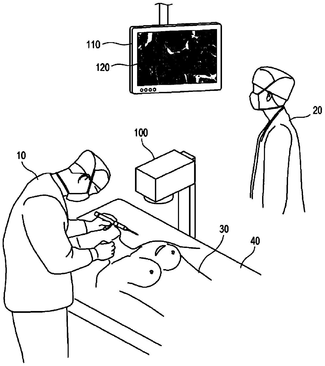
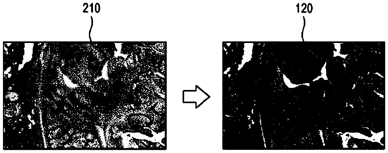
[0033] figure 1 An operating room environment is shown in which, during a medical procedure, the medical imaging apparatus 100 is running and a modified image 120 of a histological image captured by the medical imaging apparatus 100 is displayed on the display device 110 according to an embodiment of the present invention . In the operating room shown, doctors 10 and / or 20 can operate o...


PUM


Abstract

A method for processing one or more histological images captured by a medical imaging device is disclosed. In this method, the histological image is received, and target regions each of which corresponds to a candidate type of tissue are identified based on a predictive model as sociating one more sample histological images with one or more sample target histological images. One or more display characteristics associated with the identified at least one target histological image is applied to the histological image.

Description

technical field [0001] This application is based on and claims priority to U.S. Provisional Patent Application No. 62 / 461,490, filed February 21, 2017, and U.S. Provisional Patent Application No. 62 / 563,751, filed September 27, 2017, the entire contents of which are hereby incorporated by reference into this article. [0002] The present disclosure relates generally to processing histological images for display, and more particularly to generating modified images of histological images using a semi-supervised learning model. Background technique [0003] In histology, H&E (hematoxylin and eosin) stain has been widely used in medical diagnosis. For example, to examine a suspected lesion, such as cancer in a patient, a physician may obtain a sample of the suspected lesion and perform a predetermined procedure for producing an H&E stained photomicrograph. Doctors can then view the H&E-stained photomicrographs under a microscope to diagnose the patient's disease. To obtain H&...

Claims


Application Information

Patent Timeline
Patent Type & Authority Applications(China)
IPC IPC(8): G06T7/00G06T7/11G16H50/50
CPCG16H30/40G16H50/50G16H50/20G06T11/001G06T7/0014G06T2207/20081G06T2207/20084G06T2207/30024G16H10/40G06T7/0012G06T7/11G06T2210/41G16H70/00G16H30/20G06N20/00G06N3/08
Inventor 肯尼思·马克·沙利文姜镇万
Owner 株式会社高迎科技
Why Eureka
Try Eureka