Multi-channel medical imaging systems

a multi-channel, medical imaging technology, applied in the field of medical imaging systems, can solve the problems of not being suitable for open surgical applications, requiring a greater cross-sectional area for the endoscope,

Inactive Publication Date: 2016-07-21
BETH ISRAEL DEACONESS MEDICAL CENT INC
View PDF7 Cites 182 Cited by
  • Summary
  • Abstract
  • Description
  • Claims
  • Application Information

AI Technical Summary

Problems solved by technology

As a significant disadvantage, the Imaizumi system employs a number of separate cavities within an endoscopic tool for light sources and image capture, thus requiring a greater cross-sectional area for the endoscope.
As a further disadvantage, the Imaizumi patent only discloses endoscopic applications, and may not be suitable for use in open surgical applications where ambient light may extend into the excitation and / or emission wavelengths of the dye.

Method used


Image

  • Multi-channel medical imaging systems
  • Multi-channel medical imaging systems
  • Multi-channel medical imaging systems

Examples


Embodiment Construction

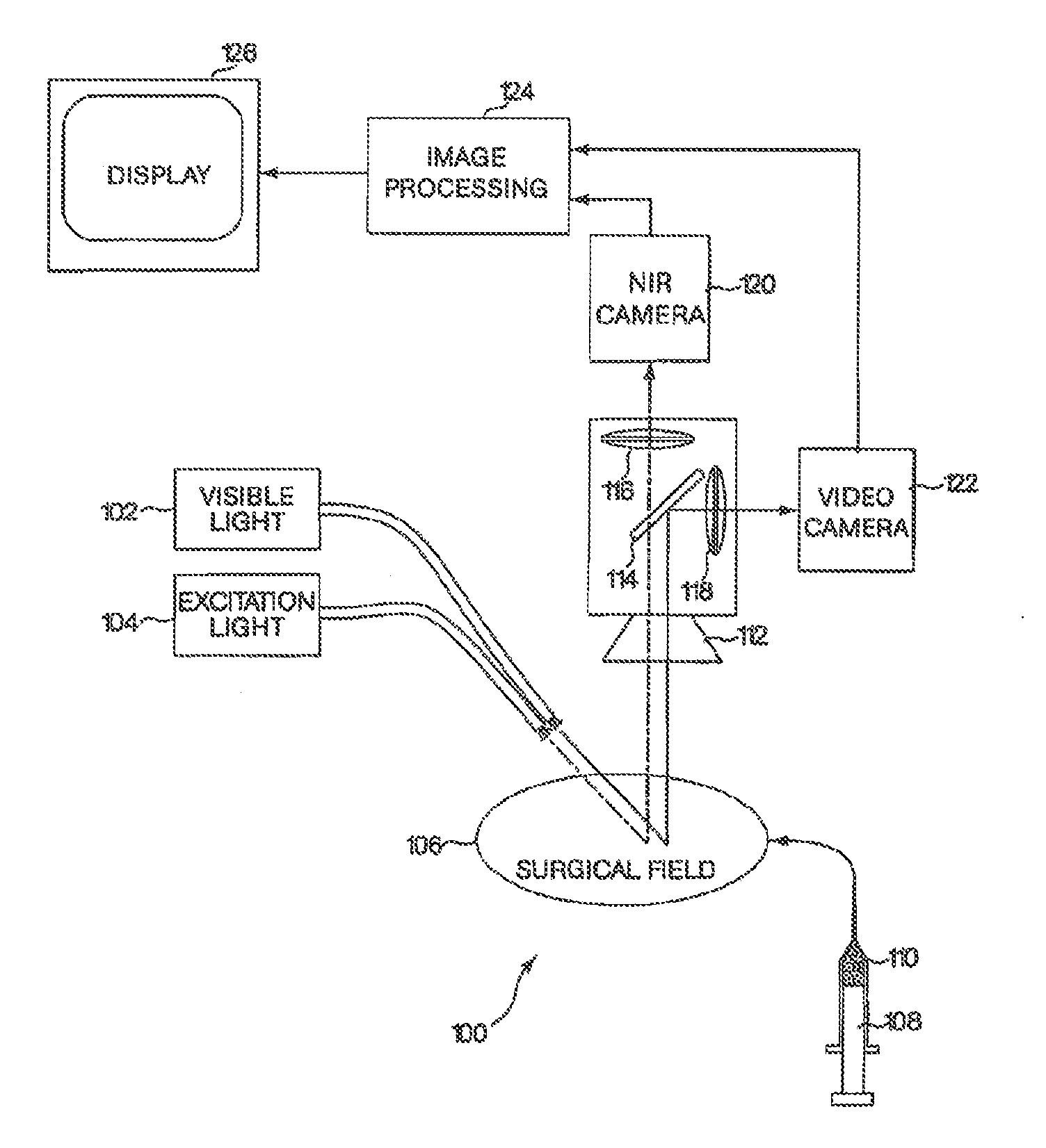
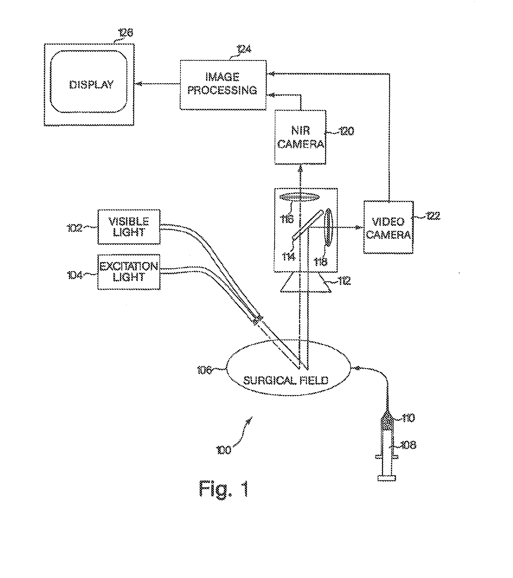
[0020]To provide an overall understanding of the invention, certain illustrative embodiments will now be described, including a system for generating superimposed circulatory and tissue images in video format. However, it will be understood that the methods and systems described herein can be suitably adapted to other medical imaging applications where visible light tissue images may be usefully displayed with diagnostic image information obtained from outside the visible light range and superimposed onto the visible light image. More generally, the methods and systems described herein may be adapted to any imaging application where a visible light image may be usefully displayed with a superimposed image captured from areas within the visible light image that are functionally marked to emit photons outside the visible light range by a dye or other material. For example, the systems and methods are applicable to a wide range of diagnostic or surgical applications where a target path...


PUM


Abstract

A medical imaging system provides simultaneous rendering of visible light and fluorescent images. The system may employ dyes in a small-molecule form that remain in a subject's blood stream for several minutes, allowing real-time imaging of the subject's circulatory system superimposed upon a conventional, visible light image of the subject. The system may provide an excitation light source to excite the fluorescent substance and a visible light source for general illumination within the same optical guide used to capture images. The system may be configured for use in open surgical procedures by providing an operating area that is closed to ambient light. The systems described herein provide two or more diagnostic imaging channels for capture of multiple, concurrent diagnostic images and may be used where a visible light image may be usefully supplemented by two or more images that are independently marked for functional interest.

Description

RELATED APPLICATIONS[0001]This application is a national phase filing under 35 U.S.C. §371 of published International Application No. PCT / US07 / 072803, filed on Jul. 3, 2007, which is incorporated herein by reference in its entirety. International Application PCT / US07 / 72803 was published in English as Publication No. WO 2008 / 042486. International Application PCT / US07 / 72803 claims priority to U.S. Prov. App. No. 60 / 818,365, filed on Jul. 3, 2006, the entire content of which is incorporated herein by reference. This application is also a continuation-in-part of U.S. patent application Ser. No. 10 / 507,253, filed on Sep. 10, 2004, incorporated herein by reference in its entirety. U.S. application Ser. No. 10 / 507,253 was filed as a U.S. national phase filing of International App. No. PCT / US03 / 07596, filed on Mar. 11, 2003, which claims priority to U.S. Prov. App. No. 60 / 363,413, filed on Mar. 12, 2002. The benefit of the foregoing applications is claimed herein to the extent permitted by ...

Claims


Application Information

Patent Timeline
Patent Type & Authority Applications(United States)
IPC IPC(8): A61B5/00A61B1/313A61B90/00A61B1/04
CPCA61B5/0071A61B5/0084A61B90/39A61B1/3132A61B90/361A61B1/043A61B1/313A61B1/0005A61B5/0035
Inventor FRANGIONI, JOHN V.
Owner BETH ISRAEL DEACONESS MEDICAL CENT INC
Why Eureka
Try Eureka