System for two-dimensional and three-dimensional imaging of tubular structures in the human body

a tubular structure and two-dimensional imaging technology, applied in the field of two-dimensional and three-dimensional imaging of tubular structures in the human body, can solve the problems of poor patient acceptance, poor compliance, hampered colorectal screening and preventive efforts, etc., to enhance the ability of computed tomographic colography (ctc), shorten the interpretation time, and reduce the interpretation time of scanned image data

Inactive Publication Date: 2005-08-09
MAYO FOUND FOR MEDICAL EDUCATION & RES
View PDF19 Cites 180 Cited by
  • Summary
  • Abstract
  • Description
  • Claims
  • Application Information

AI Technical Summary

Benefits of technology

[0024]The present invention also provides an alternative method, system, and article of manufacture for reduction in interpretation time of scanned image data involved in the production of reformatted 2D images at cross sections and orthogonal, etc. One of the ways is to display synchronously both prone and supine scanned images of the colon. This approach enhances the ability of Computed Tomographic Colography (CTC) that combines with advantages of prone and supine views without the added interpretation times of reviewing two separate scans. Simultaneous display of synchronized (anatomically registered) views of the colon eliminates the need for two separate readings of the colon and shortens interpretation time. In one embodiment of the present invention, the anatomic levels are indexed to match each other and are advanced synchronously as a radiologist interprets the data set. Axial, reformatted 2D and 3D images are displayed and simultaneously updated for both prone and supine images on the same computer screen. The colon only needs to be reviewed once, with the diagnostic benefit of both supine and prone scans. In many cases, the two scans can be interpreted nearly as quickly as one scan.
[0025]Accordingly, the present invention is also a computed tomographic colography (CTC) system having such improved interpretation tools which provide synchronously interactive montage of images. The present system displays several distinct types of images. These include volume rendered endoluminal perspective (virtual colonoscopic views), transcolonic sections, navigational scout images showing the entire colon, and “mathematically unraveled” views of the colon. The region of the screen where the scouts and small axial, sagittal, and coronal views are normally displayed can alter

Problems solved by technology

Unfortunately, widespread colorectal screening and preventive efforts are hampered by several practical impediments, including limited resources, methodologic inadequacies, and poor patient acceptance leading to poor compliance.
In addition, a fecal occult blood test (FOBT) fails to detect the majority of cancers and pre cancerous polyps.
The accuracy of barium enema varies among centers and is therefore not always reliable.
However, direct interpretation of the cross-sectional images is difficult because a scan of the colon consists of several hundred

Method used


Image

  • System for two-dimensional and three-dimensional imaging of tubular structures in the human body
  • System for two-dimensional and three-dimensional imaging of tubular structures in the human body
  • System for two-dimensional and three-dimensional imaging of tubular structures in the human body

Examples


Embodiment Construction

[0047]In the following detailed description of the preferred embodiments, reference is made to the accompanying drawings which form a part hereof, and in which is shown by way of illustration specific embodiments in which the invention may be practiced. It is to be understood that other embodiments may be utilized and structural changes may be made without departing from the scope of the present invention.

Glossary

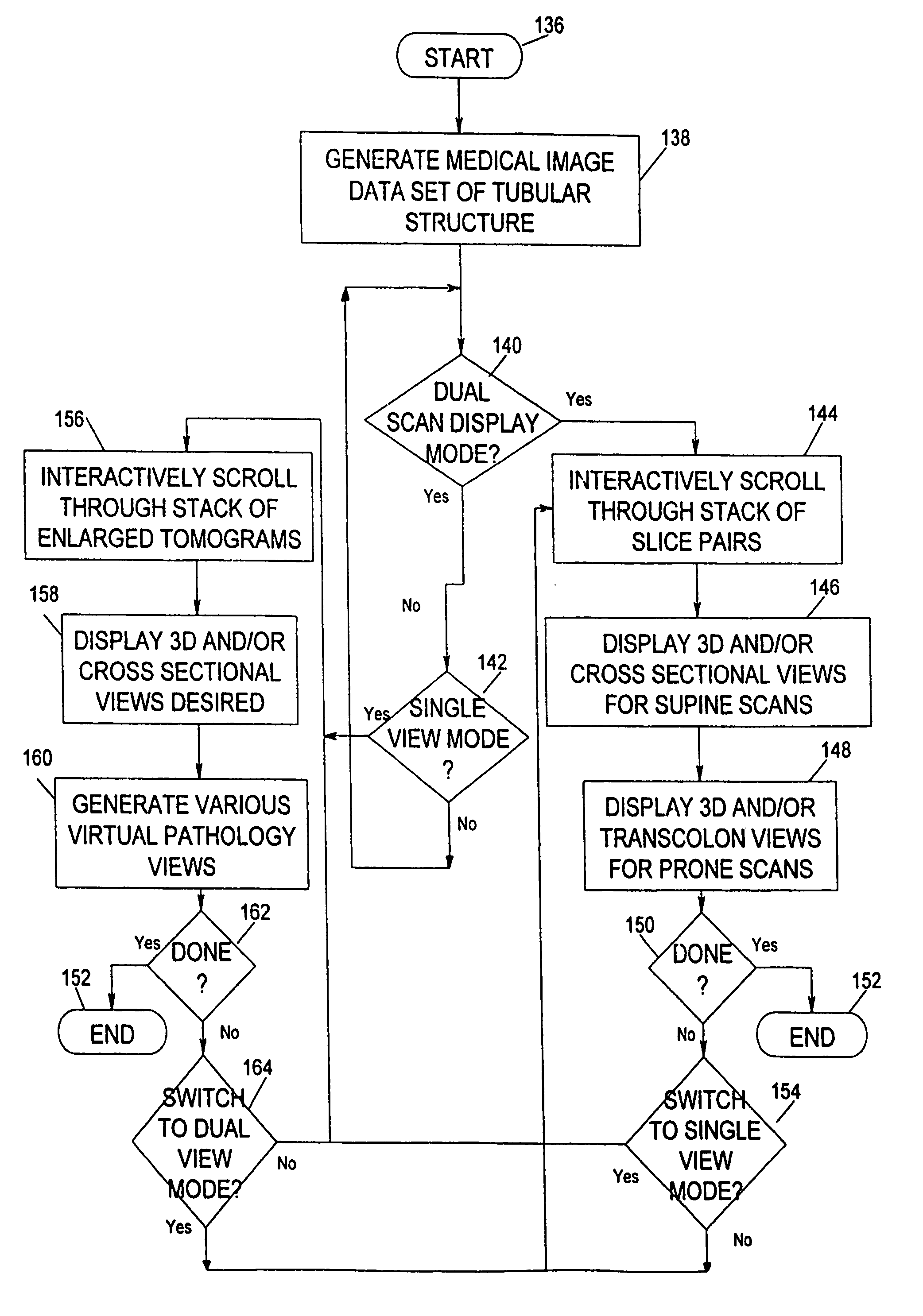
[0048]The following terms and phrases are used throughout the application.[0049]1. Computed tomographic colonography (CTC). A technique combining helical or volumetric CT imaging of the colon with imaging software to produce reformatted 2D and 3D images of the colon.[0050]2. Reformatted 2D images. Images which are oriented at cross section and two orthogonal angles to the colon midline.[0051]3. 3D intraluminal image. An image which simulates an endoscopic view of the colon. The outside of the colon is not visualized.[0052]4. 2D axial image. The original image produced by th...


PUM


Abstract

This invention is a system, method, and article of manufacture for imaging tubular structures of the human body, such as the digestive tract of a living person, with a medical imaging device such as a computed tomography (CT) scanner and a computer work station. The system comprises receiving a first image data set representative of a portion of the colon in a prone position and a second image data set representative of a portion of the colon in a supine position, at a series of viewpoints. At each of the viewpoints, an image is generated of the colon in the prone and supine positions. The prone and supine images of the colon are simultaneously displayed on a screen display in a dual view mode.

Description

FIELD OF THE INVENTION[0001]The present invention generally relates to anatomical modeling of the human body with a computer, and more particularly to a computerized system for analyzing tubular structures of the human body such as the digestive tract of a living person for detecting colorectal polyps.BACKGROUND OF THE INVENTION[0002]In industrialized nations, colorectal cancer is the second leading cause of deaths from malignancy. In the United States, almost 150,000 people are found to have colon cancer annually and it causes approximately 60,000 deaths annually. Only lung cancer causes more deaths. Colon cancers are preventable because they usually begin as benign polyps which grow slowly for five to ten years before becoming cancerous. If these polyps are detected and removed, the risk of developing colon cancer is greatly reduced.[0003]Unfortunately, widespread colorectal screening and preventive efforts are hampered by several practical impediments, including limited resources...

Claims


Application Information

Patent Timeline
IPC IPC(8): A61B5/05G06T7/60G06T15/08
CPCG06T7/606G06T15/08G06T19/003G06T2207/30028G06T2207/30172G06T2210/41Y10S128/92G06T2219/021G06T2219/028G06T7/66
Inventor JOHNSON, CHARLES D.REED, JUDD E.
Owner MAYO FOUND FOR MEDICAL EDUCATION & RES
Why Eureka
Try Eureka