Minimally invasive fixation device for tibial intercondylar spine fracture

A technology of fixation device and intercondylar spine, which is applied in the direction of fixator, internal fixator, internal bone synthesis, etc., can solve the problems that are not easy to carry out extensively, difficult to perform surgery, etc., achieve good promotion and use value, avoid large surgical trauma, and be clinically The effect of easy operation

Pending Publication Date: 2019-11-05
THE THIRD HOSPITAL OF HEBEI MEDICAL UNIV
View PDF0 Cites 0 Cited by
  • Summary
  • Abstract
  • Description
  • Claims
  • Application Information

AI Technical Summary

Problems solved by technology

[0004] The technical problem to be solved by the present invention is to provide a minimally invasive fixation device for tibial intercondylar spine fractures, through which the minimally invasive fixation device can fix tibial intercondylar spine fractures, avoiding surgical complications caused by existing open reduction and internal fixation methods. The shortcomings of large trauma, large damage, and possible complications such as infection and joint stiffness have also overcome the shortcomings of arthroscopic reduction and internal fixation, which are difficult to implement and are not easy to carry out widely. Fixed new technical support

Method used


Image

  • Minimally invasive fixation device for tibial intercondylar spine fracture
  • Minimally invasive fixation device for tibial intercondylar spine fracture
  • Minimally invasive fixation device for tibial intercondylar spine fracture

Examples


Embodiment Construction

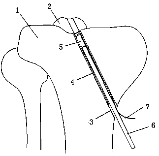
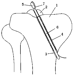
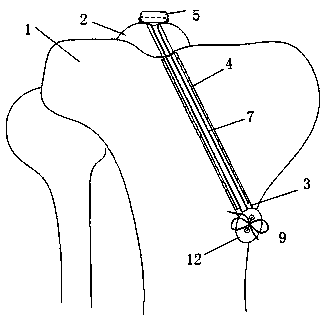
[0025] The present invention is made up of hollow sleeve 4, fixer, push rod 6, traction rope 7, and hollow sleeve 4 extends into tibial end face from the side of tibia 1, and fixer reaches tibia from hollow sleeve 4 under the push of push rod 6 On the end face, the fixing part passes through the intercondylar spinous fracture fragment 2 and reaches above the intercondylar spinous fracture fragment 2, pulls the traction rope 7 to press the fixing part above the intercondylar spinous fracture fragment 2, and tightens and fixes it from the bottom of the hollow sleeve 4 The traction rope 7 makes the fixing member fix the intercondylar spinous bone fragment 2 .

[0026] Shown in the figure, the present invention needs to punch on the tibia 1, the upper end of the fixed socket 3 of the tibia 1 is positioned at the tibial end surface below the intercondylar spinous fracture block 2 of the tibia 1, and the lower end of the fixed socket 3 is positioned at the tibial tuberosity below On...


PUM


Abstract

The invention relates to a minimally invasive fixation device for tibial intercondylar spine fracture, belongs to the technical field of orthopedic medical devices and is used for minimally invasive fixation of tibial intercondylar spine fracture. The technical scheme comprises that a hollow sleeve is inserted into a fixed insert hole of tibia, and the fixed insert hole is located between the tibia end face and the lower part of a tibia nodule; a fixed part is located at the upper end of the hollow sleeve; a push rod is located behind the fixed part, the upper end of the push rod presses against the lower end of the fixed part, the outer wall of the fixed part and the push rod is in sliding fit with the inner wall of the hollow sleeve, and the fixed part is connected with the upper end ofa traction rope; the lower end of the traction rope extends backward and out of the fixed insert hole of the tibia along the hollow sleeve. The minimally invasive fixation device has the advantages ofsimple structure, convenient clinical operation and simple operation process, can fix the tibial intercondylar spine fracture by minimal invasion, avoids the disadvantages of large surgical trauma and large damage caused by an open reduction and internal fixation method, overcomes the defects of difficult implementation of reduction and internal fixation surgery under arthroscope, and is the progress of minimally invasive surgery in the orthopedic medical technology.

Description

technical field [0001] The invention relates to a device for minimally invasive fixation of tibial intercondylar spine fractures, belonging to the technical field of orthopedic medical devices. Background technique [0002] At present, the incidence rate of tibial intercondylar spine fracture is about 3 / 100,000, and it is on the rise with the increase of traffic accidents and people's participation in sports. Tibial intercondylar spine fractures were first reported by Poncet in 1875. They are mostly caused by traffic accidents and sports injuries. The common injury mechanism is rotational stress combined with hyperextension injury. Tibial intercondylar spine fractures are intra-articular injuries. The clinically used classification system for intercondylar spine fractures was proposed by Meyers in 1959. This analysis system divides such fractures into 3 types according to the degree of displacement: Type I Intercondylar spine fractures are non-displaced fractures, type II i...

Claims


Application Information

Patent Timeline
Patent Type & Authority Applications(China)
IPC IPC(8): A61B17/68A61B17/84
CPCA61B17/683A61B17/685A61B17/846
Inventor 张英泽郑占乐于贤刘欢周汇霖
Owner THE THIRD HOSPITAL OF HEBEI MEDICAL UNIV
Why Eureka
Try Eureka