A CNN-based periocular organ segmentation method, device and storage medium

A technology around organs and eyes, which is applied in the fields of medical imaging and computers, can solve problems such as poor robustness, poor effect, and tediousness, and achieve the effects of fast speed, accurate outline, and reduced workload

Active Publication Date: 2022-08-05
BEIJING LINKING MEDICAL TECH CO LTD
View PDF10 Cites 0 Cited by
  • Summary
  • Abstract
  • Description
  • Claims
  • Application Information

AI Technical Summary

Problems solved by technology

The entire segmentation process requires multiple binary classification networks, the segmentation effect is not good, and the eye organs need to be post-processed through the conditional random field, so this method is not only cumbersome, but also poor in robustness

Method used


Image

  • A CNN-based periocular organ segmentation method, device and storage medium
  • A CNN-based periocular organ segmentation method, device and storage medium
  • A CNN-based periocular organ segmentation method, device and storage medium

Examples


Embodiment Construction

[0045] The present invention will be further described below in conjunction with the accompanying drawings and embodiments.

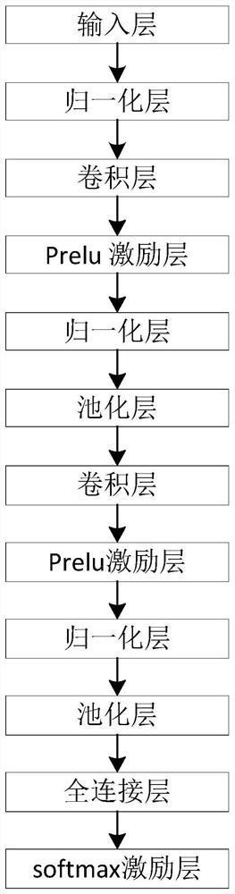
[0046] A method for accurate segmentation of periocular organs based on convolutional neural networks, wherein the periocular organs include eyes, lens, optic nerve, and pituitary gland, suitable for execution in a computing device, comprising the following steps (such as Image 6 shown):

[0047] (1) preprocessing 110 the medical image to be segmented and the medical image used as training data;

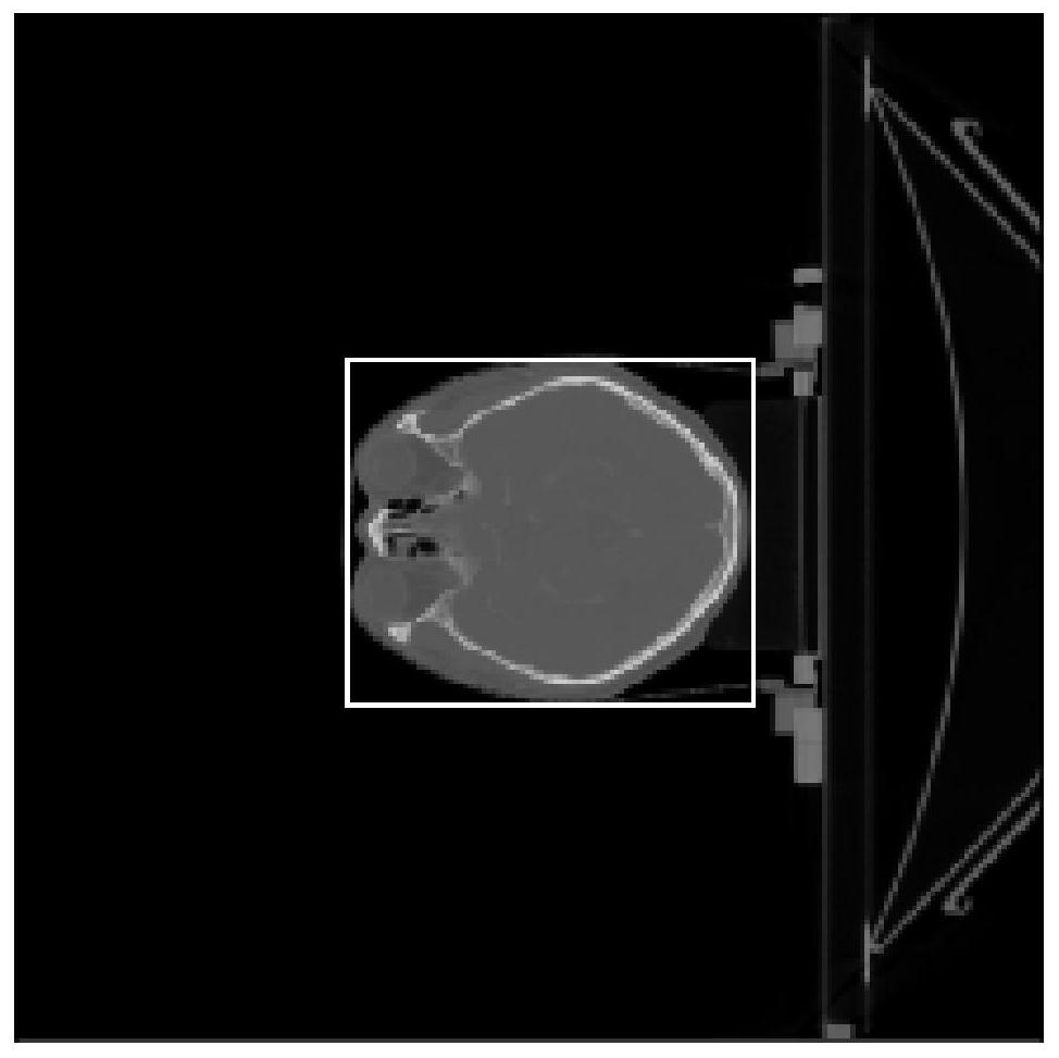
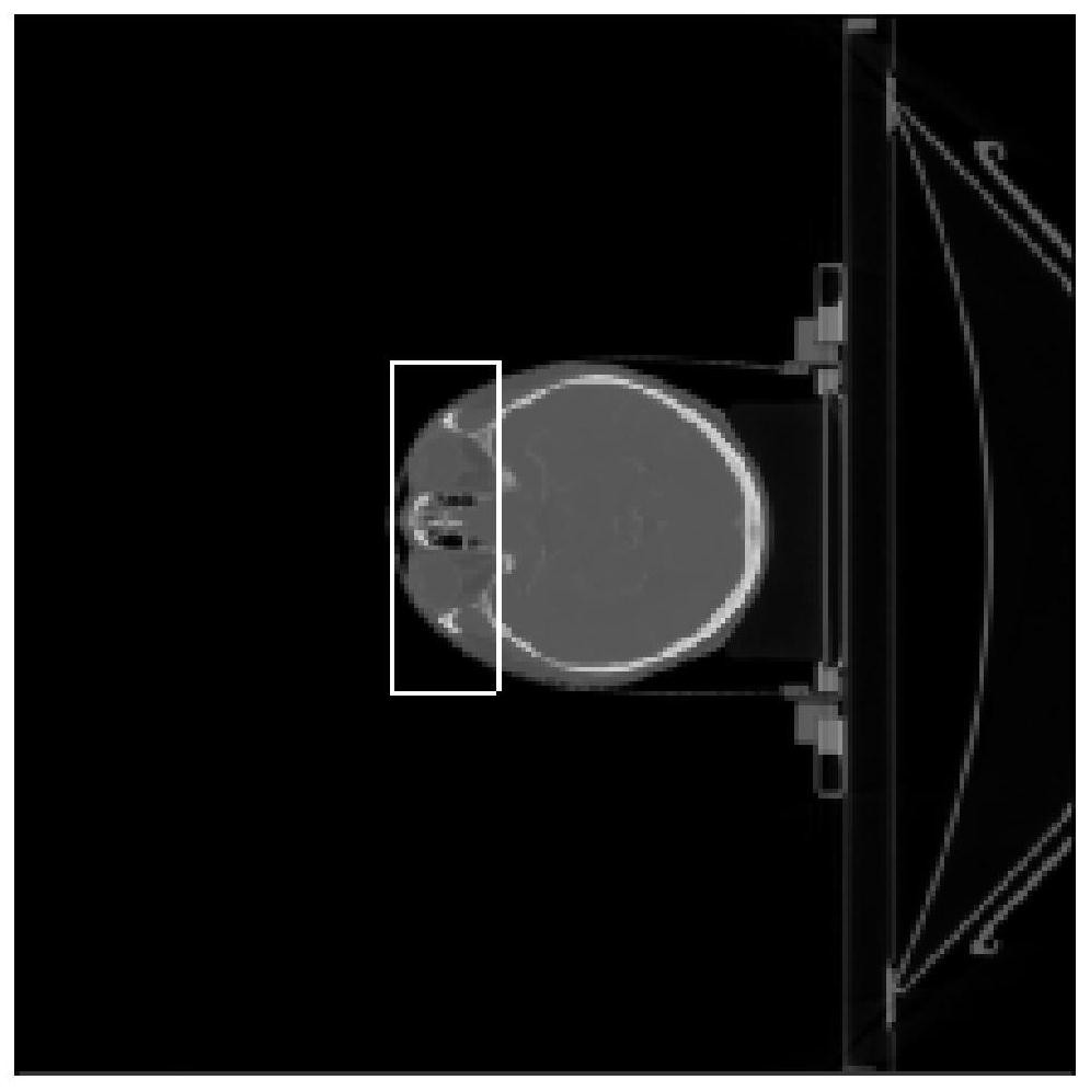
[0048] Further preferably, in this embodiment, the medical image may be selected from CT images, nuclear magnetic resonance images, PET images, ultrasound images, and the like.

[0049] Among them, the preprocessing is to eliminate the influence of metal artifacts through threshold processing. For example, metal artifacts will occur in CT images of patients with metal dentures. This is because the pixel value of dentures is much higher than that of human tis...


PUM


Abstract

The invention belongs to the technical fields of medical imaging and computers, and relates to a method, equipment and storage medium for segmenting periocular organs based on a convolutional neural network. The method includes the following steps: inputting a medical image to be segmented into a trained convolutional neural network In the two-class network, obtain the cross section containing the eye in the medical image; locate the approximate area of ​​the eye in the cross section of the eye: after segmenting the head, find the center of the head in the cross section of the eye; according to the anatomical size of the human body, locate the approximate eye through the center of the head The trained convolutional neural network for delineating the eyeball will segment the eye at the approximate position of the eye to delineate the eyeball; according to the position of the eye and the anatomy of the human body, locate the lens, optic nerve, and pituitary respectively; and then pass the corresponding volume The integrated neural network outlines the lens, optic nerve, and pituitary gland respectively.

Description

technical field [0001] The invention belongs to the field of medical imaging and computer technology, and relates to a method, device and storage medium for segmentation of periocular organs (including eye, lens, optic nerve and pituitary gland) based on convolutional neural network (CNN). Background technique [0002] Delineation of OARs (organs at risk) plays a key role in radiotherapy planning, and now OARs are mostly delineated manually by doctors and assisted by registration. For example, it takes about 3 to 4 hours to delineate each set of CT, and may need to be modified, which not only delays the patient's treatment time, but also has a large workload for doctors. [0003] Now doctors use registration to delineate OARs. However, the registration algorithm not only takes a long time, but also has unstable effect, and requires a lot of modification by doctors. With the development of artificial intelligence, deep learning is gradually applied to the segmentation method...

Claims


Application Information

Patent Timeline
Patent Type & Authority Patents(China)
IPC IPC(8): G06T7/11G06T7/136
CPCG06T7/11G06T7/136G06T2207/10081G06T2207/20081G06T2207/20084G06T2207/30041
Inventor 胡志强孙窈崔德琪史华北
Owner BEIJING LINKING MEDICAL TECH CO LTD
Why Eureka
Try Eureka