A method for collection and contamination verification of renal pelvis urine microecological specimens

A micro-ecological and urine technology, which is applied in inoculation and ovulation diagnosis, medical science, surgery, etc., can solve the problem of the lack of a collection method for obtaining renal pelvis urine micro-ecological samples, and achieve the effect of accurate judgment and high sample accuracy.

Active Publication Date: 2022-03-01
JIANGNAN UNIV
View PDF0 Cites 0 Cited by
  • Summary
  • Abstract
  • Description
  • Claims
  • Application Information

AI Technical Summary

Problems solved by technology

However, there are currently no collection methods to obtain truly representative renal pelvis urine microecological specimens

Method used


Image

  • A method for collection and contamination verification of renal pelvis urine microecological specimens
  • A method for collection and contamination verification of renal pelvis urine microecological specimens
  • A method for collection and contamination verification of renal pelvis urine microecological specimens

Examples


Embodiment 1

[0029] Collection methods of renal pelvis urine microecological specimens

[0030] 1. After the patient loses consciousness under general anesthesia, use povidone iodine to disinfect the patient's perineum and external urethral opening;

[0031] 2. Insert the ureteroscope into the bladder, take out 3mL of urine, and mark it as "bladder";

[0032] 3. Use a ureteroscope to empty all the remaining urine in the bladder;

[0033] 4. Pour iodophor into the bladder for sterilization, and observe under the ureteroscope whether the bladder is filled with iodophor, suck it out after it is full, and infuse it continuously for 3 times. The method of increasing culture is to cultivate iodine perfusate;

[0034] The expansion culture method is as follows:

[0035] (1) Inoculate 0.1mL povidone-iodine perfusion solution on sheep blood plate, chocolate plate, polymyxin plate and nalidixic acid plate respectively by 4-section line method, and inoculate them in 5% CO 2 Cultivate in an incuba...

Embodiment 2

[0042] Example 2. Verification of renal pelvis urine microecology

[0043] After extracting bacterial DNA from the samples, 16S rDNA sequencing was performed on the renal pelvis and bladder urine samples. Next, the following bioinformatics analysis methods were used to compare the differences in the urinary microecology of the renal pelvis and bladder,

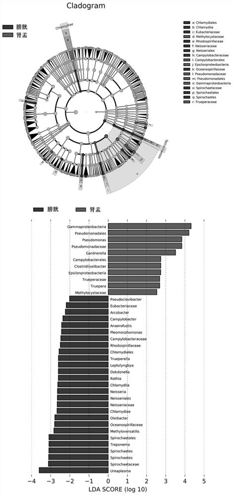
[0044] 1. LefSe analysis

[0045] According to the taxonomic composition, linear discriminant analysis is performed on the samples according to different grouping conditions to find out the communities or species that have a significant difference in the division of samples. From the analysis results, it can be seen that the microecological structure of urine from the renal pelvis is different from that of the bladder. from figure 1It can be seen that the relative abundance of Methylocystaceae, Campylobacterales, Epsilonproteobacteria, Pseudomonadales, Pseudomonadaceae, Pseudomonas, Gammaproteobacteria, Gardnerella, Clostri...


PUM


Abstract

The invention discloses a method for collecting and verifying the microecology of renal pelvis urine microecology. Further verification and comparison of the differences between the microecological structure of the renal pelvis and the microecological structure of the bladder can accurately determine whether the microecology of the renal pelvis is polluted, which is of substantial significance for the subsequent analysis of the microecology of the renal pelvis and the analysis of the relationship between the microecology of the renal pelvis and kidney diseases.

Description

technical field [0001] The invention relates to the technical field of micro-ecological specimen collection, in particular to a method for collecting micro-ecological specimens of renal pelvis urine and a method for verifying whether the renal pelvis urine specimens are polluted by bladder urine. Background technique [0002] With the development and application of high-throughput sequencing technology, the bladder, which was originally considered a sterile area, is no longer a sterile area, and the bladder microecology is independent of other organs. Like the intestinal tract, oral cavity, and vagina, the bladder not only has microecology, but its microecological structure also reflects the health status of the body. [0003] Since the microbiome exists in the bladder, there may also be a microbiome in the renal pelvis connected to it through the ureter. Since microorganisms will interact with the tissues they contact and affect the functions of cells, tissues and organs t...

Claims


Application Information

Patent Timeline
Patent Type & Authority Patents(China)
IPC IPC(8): A61B10/00
CPCA61B10/007
Inventor 柳丰萍朱升龙
Owner JIANGNAN UNIV
Why Eureka
Try Eureka