Liver segmentation method based on spatial multi-scale U-net and superpixel correction

An aspp-u-net and superpixel technology, applied in the field of liver segmentation based on spatial multi-scale U-net and superpixel correction, can solve the problem of low segmentation accuracy of fuzzy liver images, achieve smooth boundaries, accurate segmentation accuracy, easily sortable effects

Active Publication Date: 2019-11-26
SHAANXI UNIV OF SCI & TECH
View PDF2 Cites 14 Cited by
  • Summary
  • Abstract
  • Description
  • Claims
  • Application Information

AI Technical Summary

Problems solved by technology

[0006] In order to overcome the shortcomings of the above-mentioned prior art, the purpose of the present invention is to provide a liver segmentation method based on spatial multi-scale U-net and superpixel correction, which can be bet

Method used


Image

  • Liver segmentation method based on spatial multi-scale U-net and superpixel correction
  • Liver segmentation method based on spatial multi-scale U-net and superpixel correction
  • Liver segmentation method based on spatial multi-scale U-net and superpixel correction

Examples


Embodiment Construction

[0029] The present invention will be described in further detail below in conjunction with the examples.

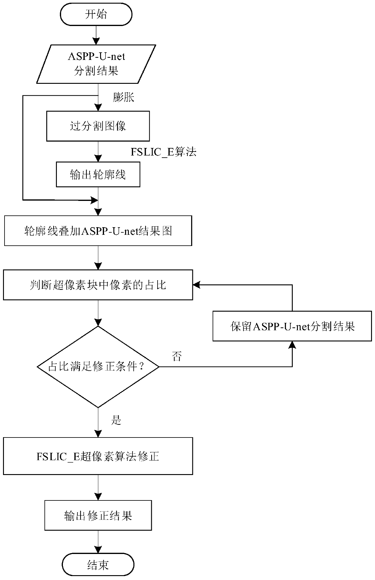
[0030] attached figure 1 It is a block diagram of the process principle of the implementation steps of the present invention. Aiming at the problem that the traditional network easily misses the position information when segmenting the image data with blurred liver boundaries, resulting in low segmentation accuracy, the present invention designs a liver segmentation method combined with superpixels and deep learning . The inventive method is specifically described as follows:

[0031] (1) Data set preprocessing: First, the W / L windowing algorithm is used to set the liver CT data to an appropriate contrast. The steps of the W / L algorithm are as follows:

[0032] (a) The formula for converting image DICOM to HU is:

[0033] HU=D*RS+RI

[0034] Wherein, HU is the output value converted from the DICOM value of the image; D is the DIOCM value of the image; RS is the readjust...


PUM


Abstract

A liver segmentation method based on spatial multi-scale U-net and superpixel correction comprises the following steps: firstly, enhancing the contrast of a liver image by using a window modulation algorithm, and suppressing noise interference through Gaussian filtering; secondly, segmenting the liver preprocessing image by using a cavity space pyramid pooling U-net model to obtain a liver preliminary segmentation result; thirdly, acquiring an over-segmentation result of the liver by utilizing a morphological expansion algorithm; and finally, correcting the liver preliminary segmentation imageby applying an FSLIC-E superpixel algorithm, and obtaining the accurate edge of the liver. According to the liver segmentation method, the respective advantages of ASPP-U-net and FSLIC_E superpixel algorithms are combined, so that a problem of relatively poor robustness of U-net for liver image segmentation is solved, and the liver segmentation method can be better applied to automatic segmentation of the liver image, so as to solve a problem of low segmentation precision of a traditional network for the blurred liver image, and realizes high segmentation precision.

Description

technical field [0001] The invention belongs to the technical field of image processing and pattern recognition, in particular to a liver segmentation method based on spatial multi-scale U-net and superpixel correction. Background technique [0002] In the initial diagnosis of liver disease, medical images have been used as the basis for doctors' diagnosis and treatment to initially judge the severity of liver disease. In order to assist doctors in diagnosing and formulating treatment plans for patients with liver diseases, it is necessary to accurately segment the liver area. At present, computer vision combined with medical imaging research has become a hotspot in the field of intelligent medical care. Liver segmentation technology based on medical imaging can obtain information such as liver size and geometric shape, thereby assisting doctors in initial diagnosis and treatment. [0003] At present, researchers have proposed a large number of liver segmentation algorithm...

Claims


Application Information

Patent Timeline
IPC IPC(8): G06T7/10G06T7/11G06T5/00
CPCG06T7/10G06T7/11G06T5/002G06T2207/30056G06T2207/10081
Inventor 雷涛连倩丁菊敏加小红刘鹏
Owner SHAANXI UNIV OF SCI & TECH
Why Eureka
Try Eureka