
Device and method for separating of partition bone and tendon interface bracket

A separation device and interface technology, applied in applications, bone implants, surgical instruments, etc., can solve the problems of tumorigenic risk of exogenous seed cells, lack of directional induced differentiation of stem cells, cumbersome operation process, etc.

Active Publication Date: 2019-12-03
XIANGYA HOSPITAL CENT SOUTH UNIV
View PDF14 Cites 1 Cited by
  • Summary
  • Abstract
  • Description
  • Claims
  • Application Information

AI Technical Summary

Problems solved by technology

The traditional bone-tendon interface regeneration strategy of tissue engineering has the following problems: 1) Existing scaffolds are only biomimetic designed from one or two aspects of morphological structure, gradient mineralization, and mechanical properties, and lack multiple biomimetic properties; 2) Existing scaffolds Most of them are made of artificial synthetic materials, and lack the characteristics of stem cell-directed differentiation induction in different areas of bone, fibrocartilage and tendon of the scaffold; 3) The introduction of exogenous seed cells has the risk of tumorigenesis, and the seed cells are loaded on the scaffold in vitro, The operation process of constructing tissue engineered grafts is cumbersome and difficult to preserve

Method used


Image

  • Device and method for separating of partition bone and tendon interface bracket
  • Device and method for separating of partition bone and tendon interface bracket
  • Device and method for separating of partition bone and tendon interface bracket

Examples


Embodiment 2

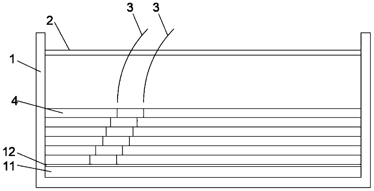
[0036] A separation method for partitioning the bone-tendon interface support, in combination with the device of embodiment 1, comprising the following steps:

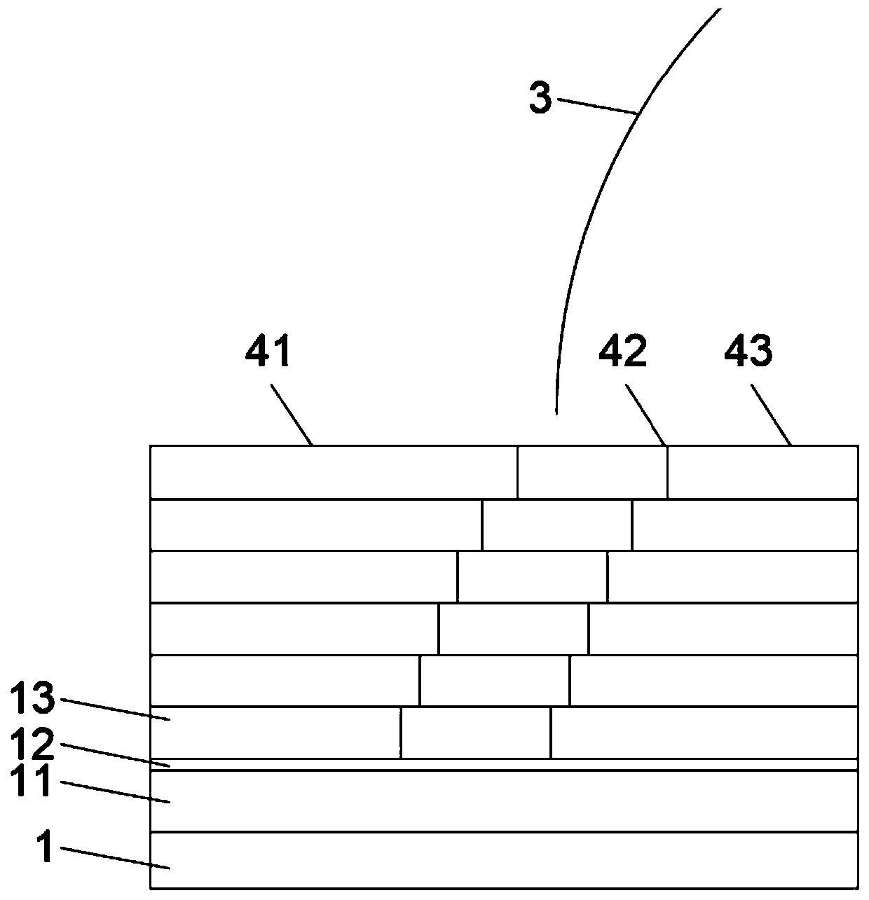
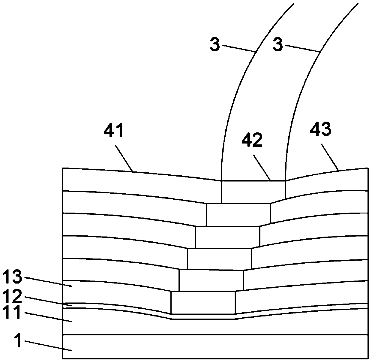
[0037] S1. Place several three-phase tissue sheets 4 on the bottom of the first cavity from top to bottom to form a three-phase tissue scaffold with several scaffold layers; wherein, each scaffold layer includes a three-phase tissue sheet 4, and the three-phase The tissue slice 4 includes a bone area 41 , a cartilage area 42 and a tendon area 43 distributed sequentially along the transverse direction. It should be understood that under normal circumstances, each scaffold layer is only composed of one three-phase structure sheet 4 (or one three-phase structure sheet 4 constitutes one scaffold layer), but other situations also fall within the protection scope of the present application.

[0038] S2. Press the partition plate 3 on the three-phase tissue sheet 4, and form a bone partition, a cartilage partition and a tendo...


PUM


Abstract

The application discloses a device and method for separating of a partition bone and tendon interface bracket. The method comprises the following steps of S1, placing a plurality of tri-phase tissue slices at the bottom of a first accommodating cavity from top to bottom, so as to form a tri-phase tissue bracket with a plurality of bracket layers; S2, pressing obstructing boards to the tri-phase tissue slices, and forming a bone obstructing region, a cartilage obstructing region and a tendon obstructing region in the first accommodating cavity through the obstructing boards; wherein the bone obstructing region, the cartilage obstructing region and the tendon obstructing region are matched with a bone region, a cartilage region and a tendon region of the tri-phase tissue bracket; and S3, adding bone growth factor fluid to the bone obstructing region, adding cartilage growth factor fluid to the cartilage obstructing region, and adding tendon growth factor fluid to the tendon region. The tri-phase tissue slices prepared through the adoption of the method disclosed by the invention have bionic characteristics, and have normal tissue patterns instead of a simple leveling structure, so that the device conforms to a physiologic structure of the human body.

Description

technical field [0001] The invention mainly relates to the defect repairing technology of the joint of bone and tendon, and in particular relates to a separation device and a separation method for partitioning the bone-tendon interface support. Background technique [0002] The following content is only the inventor's knowledge of the relevant technical field, and does not necessarily constitute prior art. [0003] The emergence and vigorous development of tissue engineering technology has encouraged clinicians and researchers to try to solve this problem through tissue engineering strategies. The traditional bone-tendon interface regeneration strategy of tissue engineering has the following problems: 1) Existing scaffolds are only biomimetic designed from one or two aspects of morphological structure, gradient mineralization, and mechanical properties, and lack multiple biomimetic properties; 2) Existing scaffolds Most of them are made of artificial synthetic materials, an...

Claims


Application Information

Patent Timeline
Patent Type & Authority Applications(China)
IPC IPC(8): A61F2/28A61B17/04
CPCA61B17/04A61F2/28A61F2002/2835
Inventor 吕红斌史强陈灿李萃李牧之胡建中
Owner XIANGYA HOSPITAL CENT SOUTH UNIV
Learn More