Automatic positioning method of standard broken-out section in ultrasonic volume images in fetuses in prenatal stage

A volumetric image, automatic positioning technology, applied in ultrasonic/sonic/infrasound image/data processing, ultrasonic/sonic/infrasonic diagnosis, ultrasonic/sonic/infrasonic Permian technology, etc. Coping with clinical images, inability to generalize standard slice positioning, etc.

Active Publication Date: 2020-01-07
深圳度影医疗科技有限公司
View PDF6 Cites 8 Cited by
  • Summary
  • Abstract
  • Description
  • Claims
  • Application Information

AI Technical Summary

Problems solved by technology

Because different parts of the fetus have different characteristics and characteristics, this type of method cannot be generalized to the standard section positioning of other parts
[0006] 2. Insufficient stability and precision of the method
Previous methods rely on tradit

Method used


Image

  • Automatic positioning method of standard broken-out section in ultrasonic volume images in fetuses in prenatal stage
  • Automatic positioning method of standard broken-out section in ultrasonic volume images in fetuses in prenatal stage
  • Automatic positioning method of standard broken-out section in ultrasonic volume images in fetuses in prenatal stage

Examples


Embodiment Construction

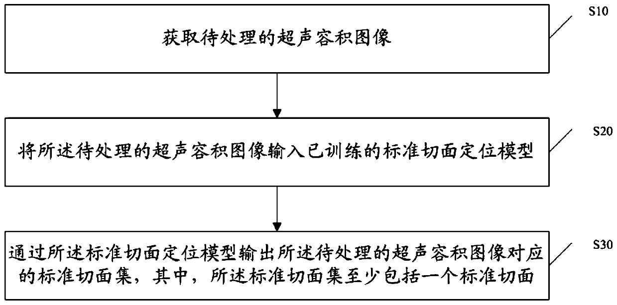
[0048] The present invention provides a method for automatic positioning of standard slices in prenatal fetal ultrasound volume images. In order to make the purpose, technical solution and effect of the present invention clearer and clearer, the present invention will be further described in detail below with reference to the accompanying drawings and examples. It should be understood that the specific embodiments described here are only used to explain the present invention, not to limit the present invention.

[0049]Those skilled in the art will understand that unless otherwise stated, the singular forms "a", "an", "said" and "the" used herein may also include plural forms. It should be further understood that the word "comprising" used in the description of the present invention refers to the presence of said features, integers, steps, operations, elements and / or components, but does not exclude the presence or addition of one or more other features, Integers, steps, opera...


PUM


Abstract

The invention discloses an automatic positioning method of standard broken-out section in ultrasonic volume images in fetuses in a prenatal stage. The method comprises the steps of obtaining ultrasonic volume images to be treated; inputting the ultrasonic volume images to be treated in a trained standard broken-out section positioning model; and outputting a standard broken-out section set corresponding to the ultrasonic volume images to be treated by a standard broken-out section positioning model. Through the standard broken-out section positioning model, the standard broken-out section setof the ultrasonic volume images can be positioned, the characteristic information of the ultrasonic volume images of three-dimensional fetuses can be sufficiently used, and the standard broken-out section set of the ultrasonic volume images can be quickly and accurately determined, so that automatic positioning of the standard broken-out section of the ultrasonic volume images can be realized.

Description

technical field [0001] The invention relates to the field of ultrasound technology, in particular to an automatic positioning method for a standard slice in antenatal fetal ultrasound volume images. Background technique [0002] Ultrasound technology is widely used in various non-invasive examinations due to its safety and rapidity, especially the non-radiation characteristics make it play a pivotal role in prenatal examinations. With the improvement of medical and health level and people's health awareness, detailed prenatal examination has become a necessary work for pregnant women. With the help of advanced ultrasound equipment, doctors can carry out relevant biological measurements on fetuses in real time and accurately, which has made great contributions to reducing the deformity rate and mortality of infants. [0003] In traditional ultrasound diagnosis, doctors perform various prenatal measurements based on the combination of two-dimensional ultrasound images and the...

Claims


Application Information

Patent Timeline
IPC IPC(8): A61B8/08
CPCA61B8/0866A61B8/5207
Inventor 杨鑫高睿钱继宽
Owner 深圳度影医疗科技有限公司
Why Eureka
Try Eureka