Intravoxel incoherent motion MRI 3-dimensional quantitative detection of tissue abnormality with improved data processing
A technology of data and organization, applied in the direction of image data processing, application, surgery, etc.
- Summary
- Abstract
- Description
- Claims
- Application Information
AI Technical Summary
Problems solved by technology
Method used
Image
Examples
Embodiment 1
[0052] A study was conducted using MRI data from the Shenzhen 2012 / 2013 dataset to determine the feasibility of using multi-parameter analysis to distinguish between fibrotic and normal liver tissue (the dataset is described in: PXLu et al., “Decreases inmolecular diffusion, perfusion fraction and perfusion-related diffusion infibrotic livers: a prospective clinical intravoxel incoherent motion MRimaging study, "PLoS One 9(12):e113846(2014)). The individuals included 16 individuals with normal liver (F0 fibrosis stage) and 33 individuals with hepatitis B virus and different degrees of liver fibrosis (F1-F4 fibrosis stage). According to the routine diagnosis based on histology, the fibrosis stage of each individual is determined. In this example, the labeling of the fibrosis stage follows the conventional scheme, where F0 stage indicates no fibrosis; F1 stage indicates only mild fibrosis can be seen in the hepatic portal area; F2 stage indicates that fibrosis extends from the he...
Embodiment 2
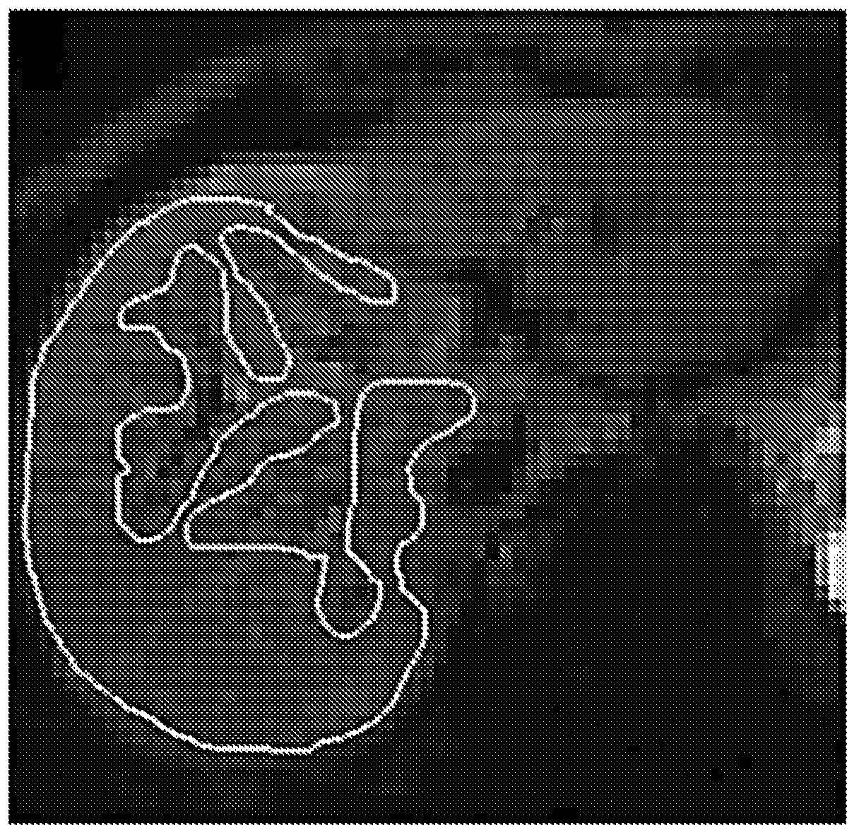
[0072] Using MRI data from the Shenzhen 2012 / 2013 dataset (see the description of Example 1 above), a study was conducted to determine the best threshold b value for distinguishing healthy and fibrotic liver tissue. The right lobe of the liver is selected for analysis, and the definition of ROI is similar to image 3 Shown. Specifically, for each individual, manually position the ROI at b=10s / mm 2 To cover most of the right liver parenchyma, while avoiding large blood vessels; the same ROI extends to include all b-value images of the individual.
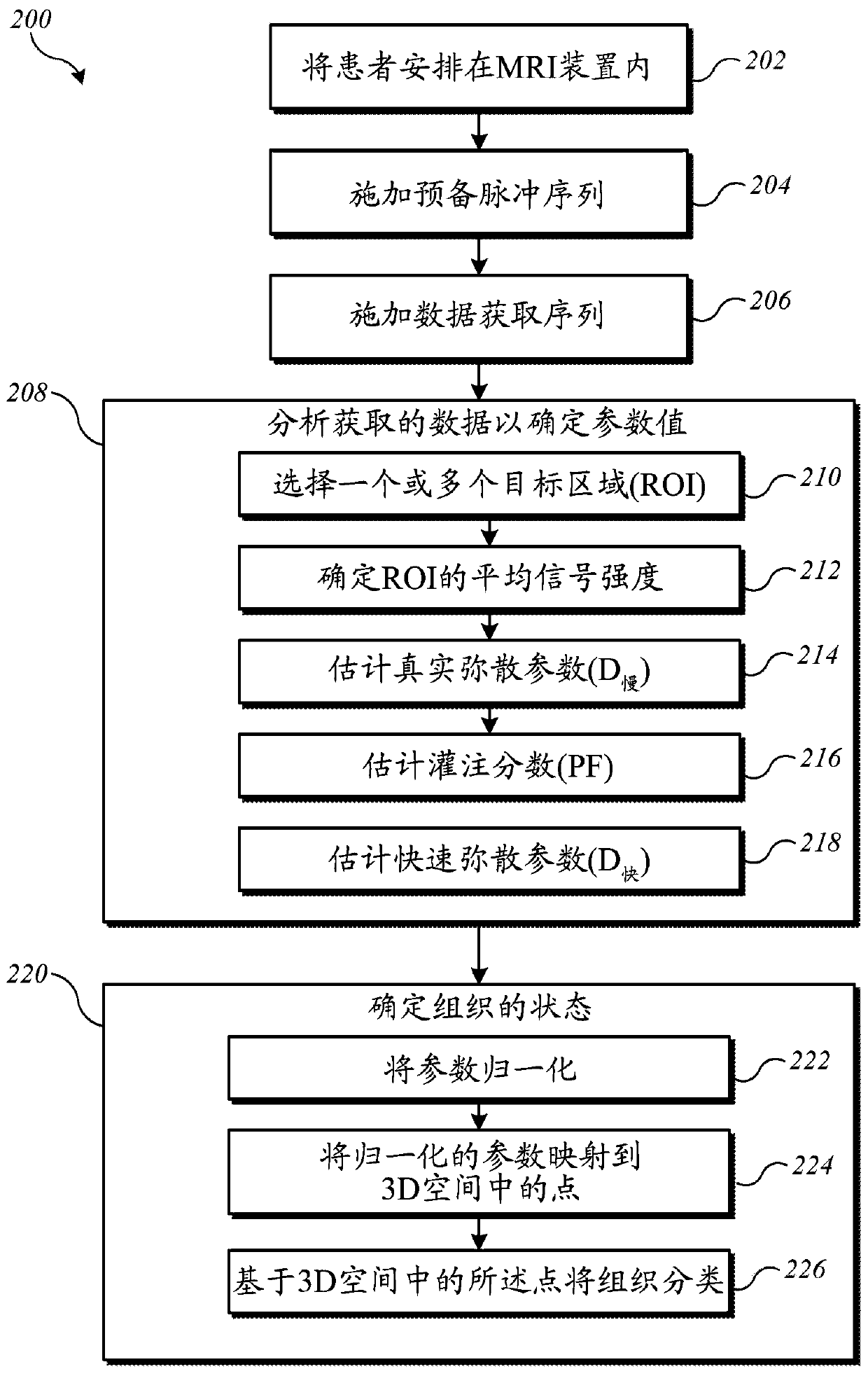
[0073] For each individual, figure 2 The analysis process of block 208 is executed 6 times, each time using a different threshold b value to determine D slow ; The threshold b value used is 40, 60, 80, 100, 150 and 200s / mm 2 . In each analysis, the images acquired with b value greater than or equal to the threshold are used to determine D slow ; Regardless of the threshold, all b values are used in the subsequent analysis phase. S...
Embodiment approach
[0100] It is believed that the embodiments described herein provide a non-invasive technique that can produce reliable diagnostic indicators of liver fibrosis, including early liver fibrosis that can only be detected by invasive techniques such as liver biopsy so far.
[0101] Although the invention has been described in terms of specific embodiments, those skilled in the art will recognize that many modifications are possible. The specific data collection sequence and analysis process can be changed. Studying a larger number of individuals with known stages of fibrosis (including healthy livers) can allow the determination of an optimized set of diagnostic parameters. In addition, it is expected that in clinical practice, the diagnostic techniques described herein will be combined with other information about the patient's condition for diagnosis.
PUM
 Abstract
Description
Claims
Application Information
 - R&D
- Intellectual Property
- Life Sciences
- Materials
- Tech Scout
- Unparalleled Data Quality
- Higher Quality Content
- 60% Fewer Hallucinations
Browse by: Latest US Patents, China's latest patents, Technical Efficacy Thesaurus, Application Domain, Technology Topic, Popular Technical Reports.
© 2025 PatSnap. All rights reserved.Legal|Privacy policy|Modern Slavery Act Transparency Statement|Sitemap|About US| Contact US: help@patsnap.com


