Fundus blood vessel segmentation method based on branch attention and multi-model fusion

A model and blood vessel technology, which is applied in the field of fundus blood vessel segmentation based on branch attention and multi-model fusion, which can solve the problems of small blood vessels that cannot be well segmented and attention loss.

Active Publication Date: 2020-04-21
北京小白世纪网络科技有限公司
View PDF9 Cites 17 Cited by
  • Summary
  • Abstract
  • Description
  • Claims
  • Application Information

AI Technical Summary

Problems solved by technology

The invention solves the problem that some small blood vessels cannot be well segmented in the fundus image blood vessel segmentation problem, so that when there are many small blood vessels in the fundus, the segmentation model and attention loss function are designed to ensure the accuracy of the segmentation during the segmentation process. , the fundus blood vessel segmentation model can notice the small blood vessels, which improves the segmentation accuracy

Method used


Image

  • Fundus blood vessel segmentation method based on branch attention and multi-model fusion
  • Fundus blood vessel segmentation method based on branch attention and multi-model fusion
  • Fundus blood vessel segmentation method based on branch attention and multi-model fusion

Examples


specific Embodiment approach

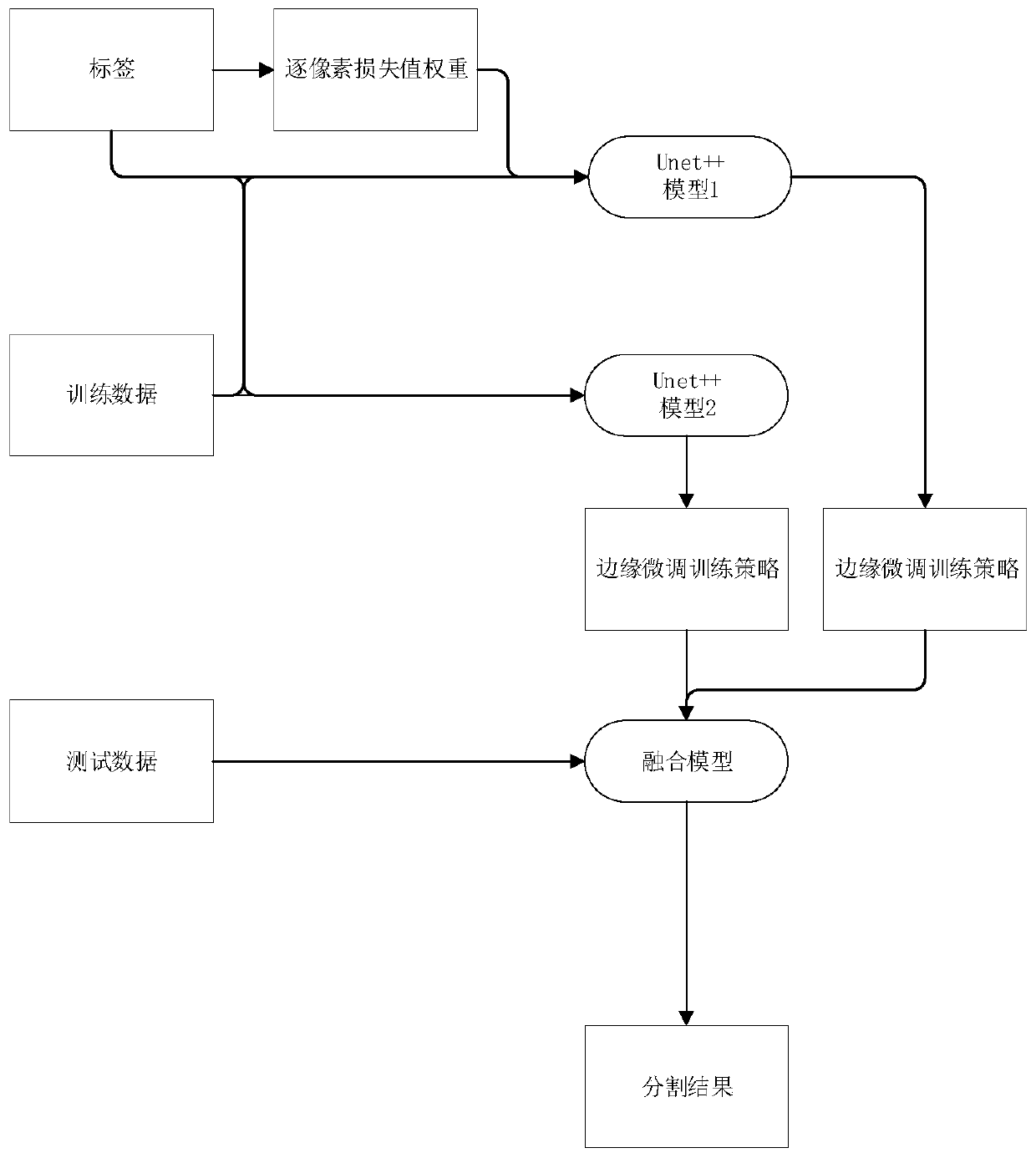
[0032] figure 1 A schematic diagram of a fundus blood vessel segmentation method based on branch attention and multi-model fusion provided by an embodiment of the present invention is shown. Its specific implementation is as follows:

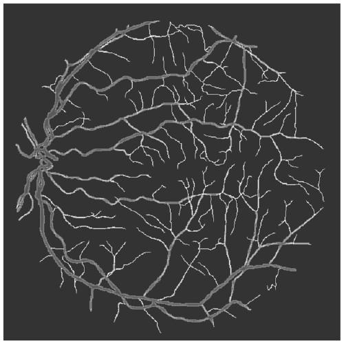
[0033] 1 fundus image enhancement

[0034]For the fundus blood vessel segmentation image, in order to better make the blood vessel pixels in the image have a higher contrast with other pixels, the image needs to be enhanced and preprocessed. In the present invention, CLAHE is used for preprocessing. At the same time, the fundus image needs to be trained Perform operations such as rotation and random cropping to expand the training data. Concrete operation is as follows, at first, record fundus image as I, note rotation operation as R (I, angle), record the random number that range is [0, N] as rand (n), CLAHE operation is CLAHE (I), and I is A fundus image, randomly cropped as CLIP(I, range), which means that the image I is randomly cropped w...


PUM


Abstract

The invention relates to a CT image organ segmentation method based on convolutional neural network multi-dimensional fusion. The method comprises the steps of S1, training a Unet++ model through weights, training data and label data which are obtained through label calculation by means of an attention loss function; S2, training the Unet++ model by utilizing the training data, the label and a binary cross entropy loss function; S3, respectively obtaining two different segmentation results by utilizing the two obtained trained Unet++ models and the to-be-segmented data; and S4, fusing the twodifferent segmentation results. According to the method, the problem that some small blood vessels cannot be well segmented in the blood vessel segmentation problem of the fundus image is solved, so that the segmentation accuracy is improved.

Description

technical field [0001] The invention relates to the technical field of medical image processing, in particular to a fundus blood vessel segmentation method based on branch attention and multi-model fusion. Background technique [0002] At present, image foreground and background segmentation technology is widely used in many real-world scenarios. How to construct an accurate and efficient image foreground and background segmentation model is the most important step in image classification technology. Existing segmentation methods are mainly based on supervised learning, that is, manually collect a large number of labeled training data of the target category, and then use these training data to build a segmentation model for the target category. [0003] Retinal vessel segmentation and description of retinal vessel morphological features, such as length, width, tortuosity, branching patterns and angles, in fundus images can be used for diagnosis, screening, treatment and eval...

Claims


Application Information

Patent Timeline
Patent Type & Authority Applications(China)
IPC IPC(8): G06T7/12G06T7/11G06T7/187G06K9/62
CPCG06T7/12G06T7/11G06T7/187G06T2207/10081G06T2207/20081G06T2207/20084G06T2207/30041G06F18/25
Inventor 杜强陈相儒郭雨晨聂方兴张兴
Owner 北京小白世纪网络科技有限公司
Why Eureka
Try Eureka