Application of PD-L1/CTLA-4 in preparation of immunosuppressant
A technology of CTLA-4 and PD-L1, which is applied in the field of biomedicine to achieve the effect of suppressing immune rejection, great application prospects, and good immunosuppressive effect
- Summary
- Abstract
- Description
- Claims
- Application Information
AI Technical Summary
Problems solved by technology
Method used
Image

Examples
Embodiment 1
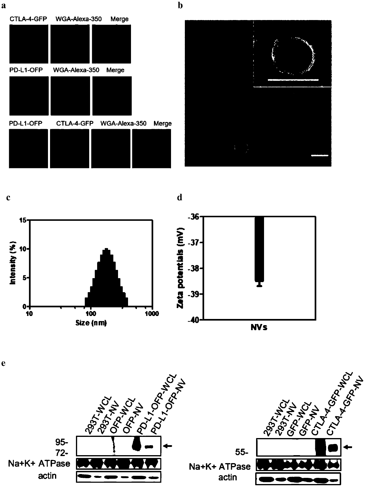
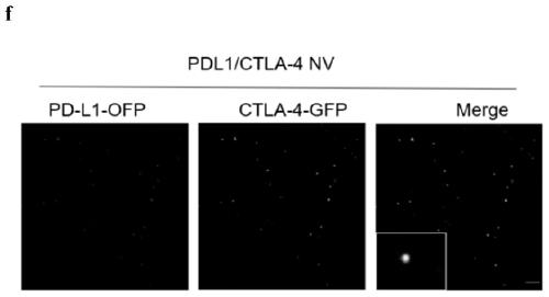
[0038] Example 1 Construction of PD-L1 / CTLA-4 membrane nanovesicles
[0039] Construction of cell lines: HEK 293T cells were transfected with packaging plasmids VSV-G, pHIV-gag-pol and targeting plasmids Lenti-PD-L1-OFP or Lenti-CTLA-4-GFP using Lipofectamine 2000 (Life Technologies). 12 and 24 hours after transfection (p.t.), the supernatant containing lentivirus was collected and filtered (0.45 μm). Then the collected virus was infected with HEK293T cells, or ASC cells and original 264.7 cells were infected with lentivirus, and screened with puromycin (2 μg / ml) to obtain stably expressed target cells. Firstly, a cell line stably expressing PD-L1-OFP was constructed, and then the cells were re-infected with CTLA-4-GFP lentivirus, and the co-infection cell line was obtained through puromycin selection.
[0040] The localization results of PD-L1 / CTLA-4 expression on the 293T cell membrane were detected by confocal laser microscopy. The result is as figure 1 As shown in a, la...
Embodiment 2
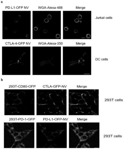
[0044] Example 2 In vitro biological behavior of PD-L1 / CTLA-4 membrane nanovesicles
[0045] (1) Nanovesicle cell binding assay: Jurkat cells were incubated with PD-L1-OFP NVs (50 μg / ml, protein weight) for 30 minutes, and centrifuged with a Ceoporep-4 centrifuge (Intech, China) to make slides. Then, wheat germ agglutinin (WGA) and Alexa-Fluor 488 conjugate were added to stain the cell membrane for 10 min, bmdc were seeded in a confocal culture dish, and DC cells were stimulated with 10 U-TNF-α. CTLA-4-OFP NVs (50 μg / ml, protein weight) were added and incubated for 30 min as previously described. Then, wheat germ agglutinin (WGA) and Alexa-Fluor 350 conjugate were added, incubated at 37° C. for 10 min, and the membrane was stained. 293T-CD80-OFP cells or 293T-PD-1-GFP cells were seeded in confocal culture dishes, respectively. CTLA-4-OFP NVs (50 μg / ml, protein weight) or PD-L1-OFP NVs (50 μg / ml, protein weight) were added to the medium and incubated for 30 minutes. Images w...
Embodiment 3
[0050] Example 3 The use of PD-L1 / CTLA-4 membrane nanovesicles in skin transplantation models
[0051] The skin graft models were established respectively. The use of experimental animals and all experiments were reviewed and approved by the Animal Ethics Committee of Sun Yat-sen Medical College, Sun Yat-sen University, with approval number 2018000577.
[0052] For skin grafting, mice were anesthetized, shaved and sterilized with 75% ethanol. The skin of male C57BL / 6 mice (0.8cm 2) were transplanted into the dorsal side of 8-week-old BALB / c mice. Fifteen recipients were randomly divided into 5 groups: PC group (normal saline, n=3), group 2 (PD-L1 NVs, 25mg / kg, n=3), group 3 (CTLA-4NVs, 25mg / kg kg, n=3), group 4 (PD-L1 / CTLA-4NVs, 25 mg / kg, n=3), NC group (autologous transplantation, n=3). Transplanted mice were dosed daily by tail vein injection for the first 3 days. After 3 days, NVs were injected every other day, and the survival curve of the mice was observed until 14 d...
PUM
| Property | Measurement | Unit |
|---|---|---|
| Particle size | aaaaa | aaaaa |
| Electric potential | aaaaa | aaaaa |
Abstract
Description
Claims
Application Information
 - R&D Engineer
- R&D Manager
- IP Professional
- Industry Leading Data Capabilities
- Powerful AI technology
- Patent DNA Extraction
Browse by: Latest US Patents, China's latest patents, Technical Efficacy Thesaurus, Application Domain, Technology Topic.
© 2024 PatSnap. All rights reserved.Legal|Privacy policy|Modern Slavery Act Transparency Statement|Sitemap