Ultrasonic image segmentation device and ultrasonic image segmentation method

An ultrasound image and ultrasound technology, applied in image analysis, image enhancement, image data processing, etc., can solve problems such as poor robustness, difficulty in distinguishing tumors, and pain in patients, to improve directness and transparency, and improve feature utilization. , optimize the effect of shallow features

Pending Publication Date: 2020-06-19
BEIHANG UNIV
View PDF6 Cites 2 Cited by
  • Summary
  • Abstract
  • Description
  • Claims
  • Application Information

AI Technical Summary

Problems solved by technology

At present, the mainstream breast cancer screening method in my country is X-ray mammography technology, but this method has certain defects: first, X-ray has certain ionizing radiation, which will cause harm to the human body; second, because this method produces two-dimensional image

Method used


Image

  • Ultrasonic image segmentation device and ultrasonic image segmentation method
  • Ultrasonic image segmentation device and ultrasonic image segmentation method
  • Ultrasonic image segmentation device and ultrasonic image segmentation method

Examples


Embodiment 1

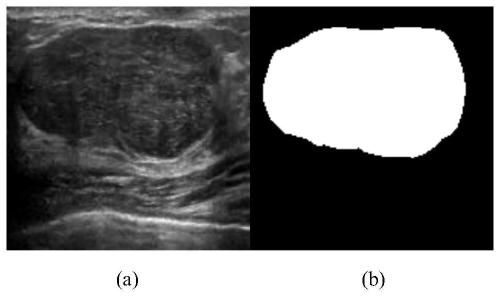
[0050] S1: Manually label breast ultrasound images. Ultrasound images and labeled images such as figure 2 (a) and figure 2 (b) shown.

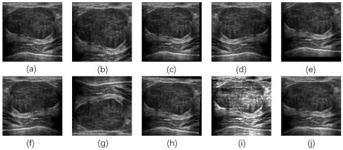
[0051] S2: Use data augmentation methods to expand the data set to avoid overfitting. The data enhancement method adopted in the present invention includes vertical and horizontal movement; vertical and horizontal flip; rotation; adding noise; zooming in and out; Various enhancement methods such as image 3 (a)- image 3 (j) shown.

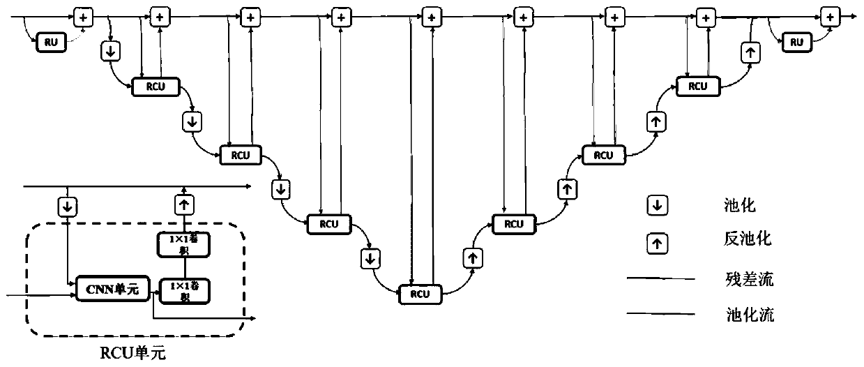
[0052] S3: Add the designed Dense module to the FRRN network to improve feature utilization and help gradient backpropagation. The module structure is as Figure 4 (a) and Figure 4 (b) shown. The input of each layer is the output of all previous layers. This design improves feature utilization and helps gradient backpropagation.

[0053] S4: Add an upsampling module based on the attention mechanism, and perform 2 ntimes the upsampling, n is the network depth at the upsampling point, and restores to t...


PUM


Abstract

The invention discloses an ultrasonic image segmentation device and an ultrasonic image segmentation method, which generate and display image data of an examination part based on volume data generatedby ultrasonic scanning of an examined object, and comprise an image enhancement module, an FRRN module and an output module which are cascaded. The technical scheme of the invention is applied to anultrasonic image segmentation task, so that the network automatically learns image features, and the segmentation precision and robustness are improved.

Description

technical field [0001] The invention belongs to the technical field of automatic biomedical ultrasonic image segmentation, and in particular relates to an ultrasonic image segmentation device and an ultrasonic image segmentation method. Background technique [0002] Today, breast cancer has become the most common cancer among women in the world. Early screening of breast cancer and timely treatment can reduce mortality. At present, the mainstream breast cancer screening method in my country is X-ray mammography technology, but this method has certain defects: first, X-ray has certain ionizing radiation, which will cause harm to the human body; second, because this method produces two-dimensional images, For dense mammary glands, it is difficult to distinguish the tumor in the image; 3. The pressing of the instrument will bring pain to the patient. Compared with X-rays, ultrasound has very little ionizing radiation, and the imaging is a three-dimensional image, and the screen...

Claims


Application Information

Patent Timeline
IPC IPC(8): G06T7/00G06T7/10G06N3/04G06N3/08
CPCG06T7/0012G06T7/10G06N3/084G06T2207/10132G06T2207/20081G06T2207/20084G06T2207/30068G06N3/045
Inventor 屈晓磊侍尧
Owner BEIHANG UNIV
Why Eureka
Try Eureka