
Ultrasonic imaging method and device and storage medium

An ultrasonic imaging method and ultrasonic imaging technology, applied in image enhancement, image analysis, image data processing, etc., can solve the problems of large differences in segmentation results and low segmentation efficiency

Pending Publication Date: 2020-06-26
SHENZHEN MINDRAY BIO MEDICAL ELECTRONICS CO LTD
View PDF4 Cites 3 Cited by
  • Summary
  • Abstract
  • Description
  • Claims
  • Application Information

AI Technical Summary

Problems solved by technology

However, in the actual segmentation process, since the segmentation is completely done manually, not only the difference in the segmentation results is large, but also the segmentation efficiency is low.

Method used


Image

  • Ultrasonic imaging method and device and storage medium
  • Ultrasonic imaging method and device and storage medium
  • Ultrasonic imaging method and device and storage medium

Examples


Embodiment Construction

[0046] In order to understand the characteristics and technical contents of the embodiments of the present application in more detail, the implementation of the embodiments of the present application will be described in detail below in conjunction with the accompanying drawings. The attached drawings are only for reference and description, and are not intended to limit the embodiments of the present application.

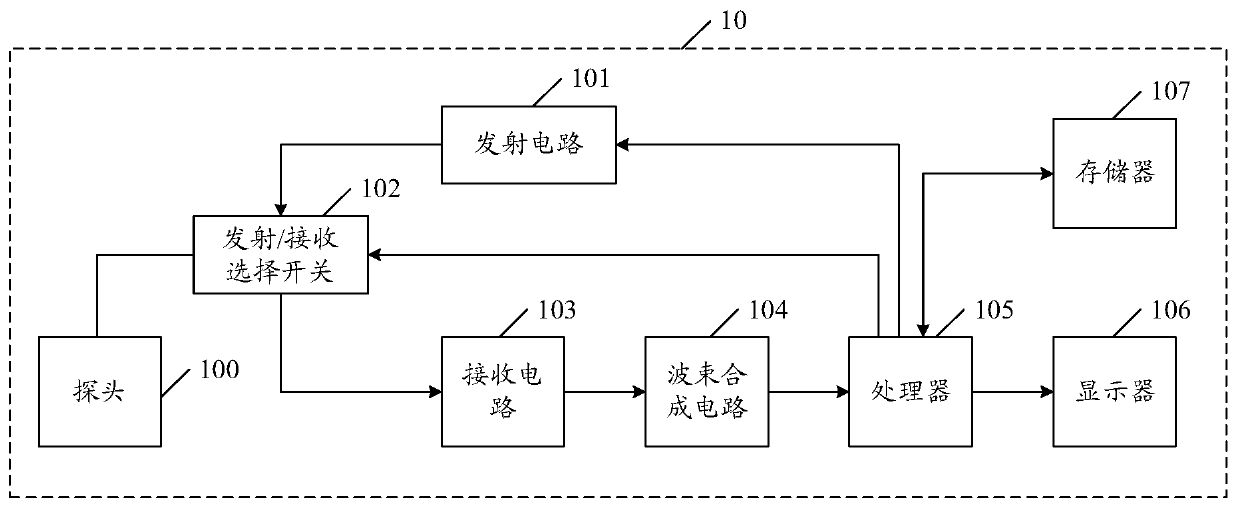
[0047] figure 1 It is a schematic structural block diagram of an ultrasonic imaging device in an embodiment of the present application. The ultrasonic imaging device 10 may include a probe 100 , a transmitting circuit 101 , a transmitting / receiving selection switch 102 , a receiving circuit 103 , a beam forming circuit 104 , a processor 105 and a display 106 . The transmitting circuit 101 can stimulate the probe 100 to transmit ultrasound to the target tissue; the receiving circuit 103 can receive the ultrasonic echo returned from the target tissue through the probe...


PUM


Abstract

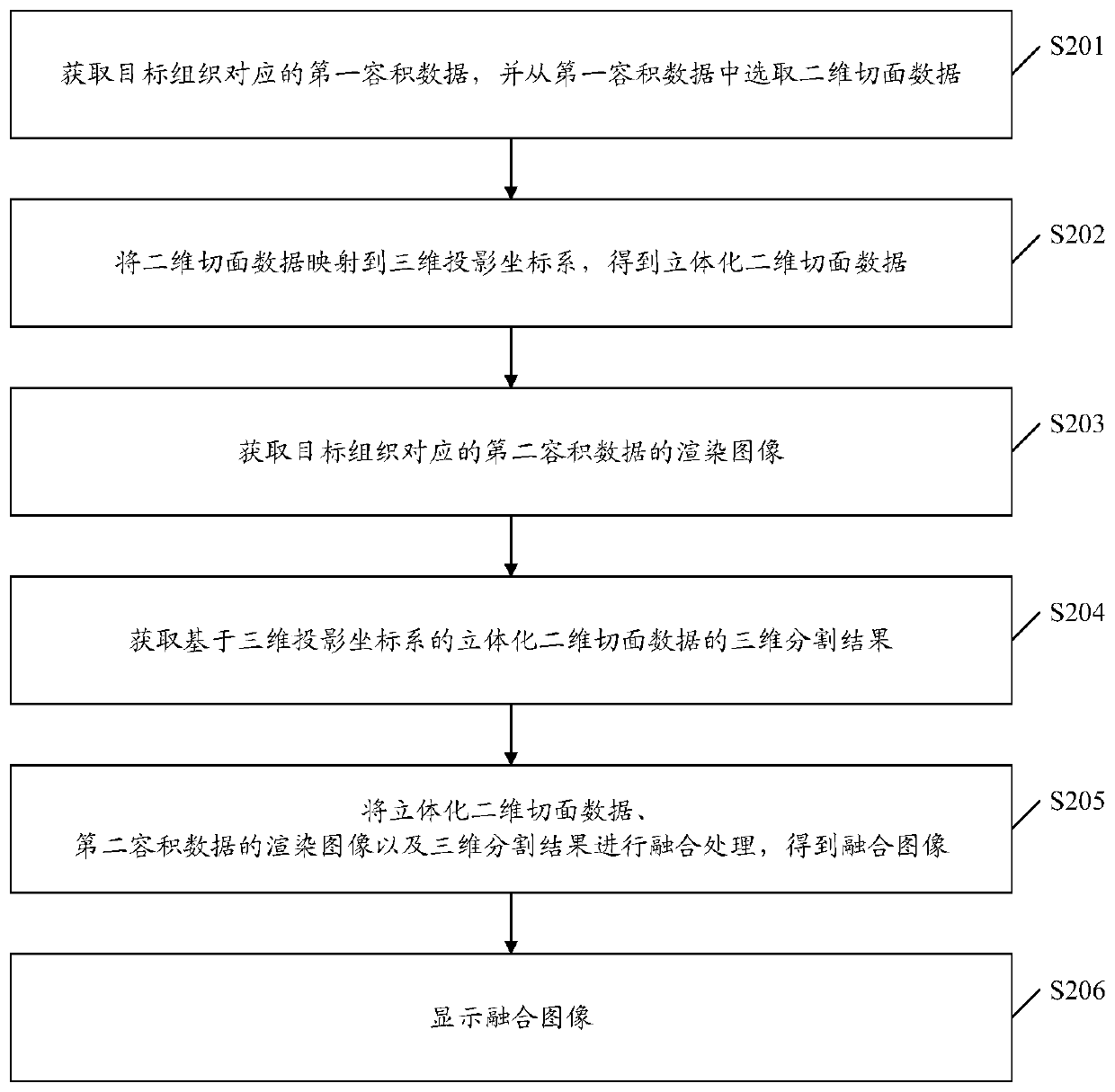
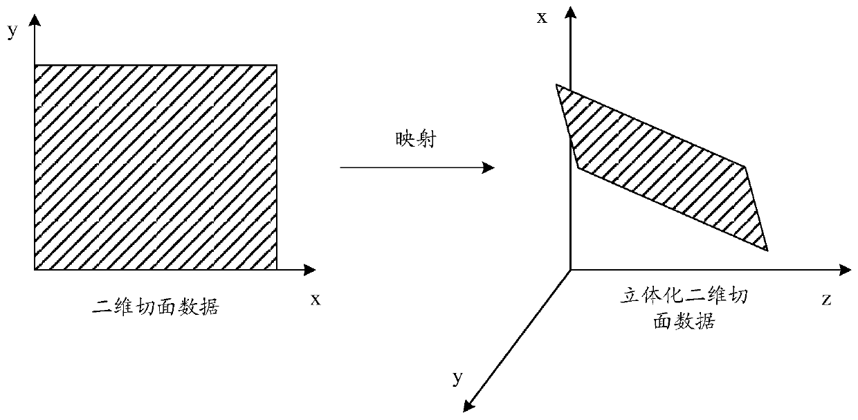
The embodiment of the invention discloses an ultrasonic imaging method, and the method comprises the steps: obtaining first volume data corresponding to a target tissue, and selecting two-dimensionalsection data from the first volume data; mapping the two-dimensional section data to a three-dimensional projection coordinate system to obtain three-dimensional two-dimensional section data; obtaining a rendering image of second volume data corresponding to the target tissue; obtaining a three-dimensional segmentation result of the three-dimensional two-dimensional section data based on a three-dimensional projection coordinate system; performing fusion processing on the three-dimensional two-dimensional section data, the rendered image of the second volume data and the three-dimensional segmentation result to obtain a fused image; and displaying fused images.

Description

technical field [0001] The present application relates to the technical field of ultrasonic imaging, and in particular to an ultrasonic imaging method, device, and storage medium. Background technique [0002] At present, ultrasonic imaging equipment can continuously collect a series of two-dimensional slice images of the target tissue, and perform three-dimensional rendering processing on the collected two-dimensional slice images, so as to obtain a three-dimensional image of the target tissue, which is convenient for doctors to more accurately and detailedly Make a medical diagnosis. [0003] Three-dimensional image segmentation refers to extracting the image information of the organ, tissue or diseased body of interest from the three-dimensional image, and then using the three-dimensional rendering technology to restore the three-dimensional structure information of the organ, tissue or diseased body of interest from the extracted image information , so that doctors can ...

Claims


Application Information

Patent Timeline
Patent Type & Authority Applications(China)
IPC IPC(8): G06T5/50G06T3/00G06T7/10
CPCG06T5/50G06T7/10G06T2207/10136G06T2207/20221G06T2207/10081G06T2207/10088G06T2207/10116G06T3/08
Inventor 王艾俊林穆清邹耀贤贾洪飞陈志杰
Owner SHENZHEN MINDRAY BIO MEDICAL ELECTRONICS CO LTD
Learn More