Automatic marking method for lesion area form in breast ultrasound contrast video

An automatic labeling, contrast-enhanced ultrasound technology, applied in the field of medical ultrasound image data processing

Active Publication Date: 2020-07-28
SOUTHWEST JIAOTONG UNIV
View PDF11 Cites 8 Cited by
  • Summary
  • Abstract
  • Description
  • Claims
  • Application Information

AI Technical Summary

Problems solved by technology

[0004] In order to solve the problems existing in the prior art, the present invention provides a method for automatically marking the shape of the lesion area in breast ultrasound contrast-enhanced video, which solves the problems mentioned in the above-mentioned background technology

Method used


Image

  • Automatic marking method for lesion area form in breast ultrasound contrast video
  • Automatic marking method for lesion area form in breast ultrasound contrast video
  • Automatic marking method for lesion area form in breast ultrasound contrast video

Examples


Embodiment Construction

[0028] The following will clearly and completely describe the technical solutions in the embodiments of the present invention with reference to the accompanying drawings in the embodiments of the present invention. Obviously, the described embodiments are only some, not all, embodiments of the present invention. Based on the embodiments of the present invention, all other embodiments obtained by persons of ordinary skill in the art without making creative efforts belong to the protection scope of the present invention.

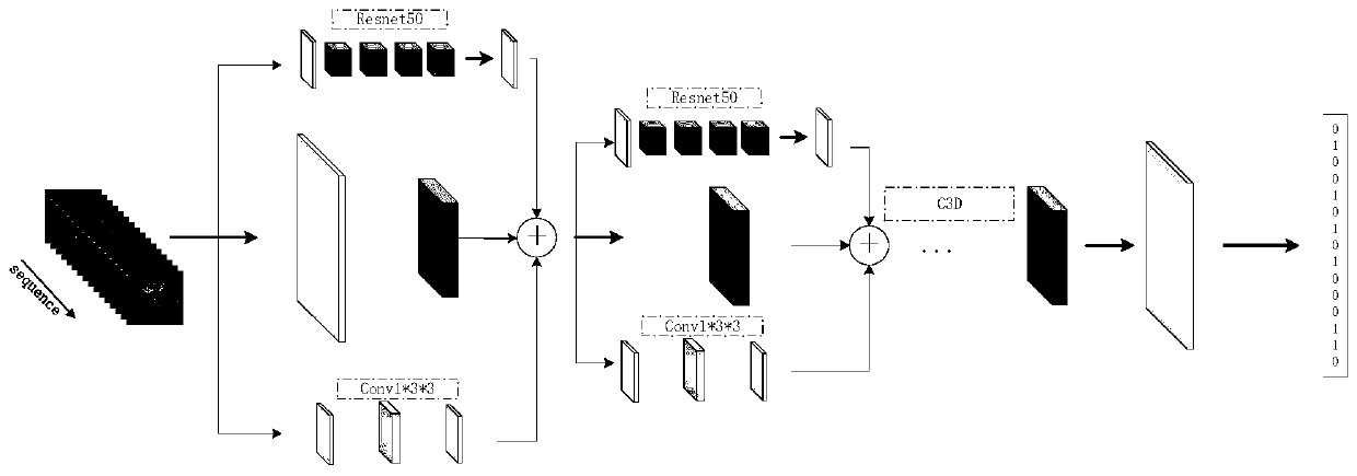
[0029] see Figure 1-5 , the present invention provides a technical solution: a method for automatically marking the shape of lesion areas in breast contrast-enhanced ultrasound videos, which uses a convolutional neural network architecture to automatically extract the feature parameters of lesion shapes in ultrasound-enhanced ultrasound videos to complete shape recognition and classification. The case data is marked with lesions, and the steps are as follows:...


PUM


Abstract

The invention discloses an automatic marking method for lesion area morphology in a breast ultrasound contrast video, and the method comprises the steps: designing an end-to-end network model structure, just transmitting to-be-recognized data to a model, enabling the model to automatically carry out the convolution operation of each frame of image, and extracting a discrimination feature of a classification basis. The focus area range does not need to be manually drawn in the whole identification process; certain lesion morphological characteristics describe contrast change under related normal tissues and contrast change of lesion tissues, such as enhanced intensity and enhanced time sequence, a convolutional neural network is used for automatically carrying out convolution calculation ona whole radiography video frame sequence, mapping data of normal tissues and lesion areas are shown through calculated characteristic values, and comparison is carried out according to network rulesto obtain a result. In addition, for morphological characteristics such as crab foot shape and enhancement sequence, the designed network is used to automatically calculate the characteristics corresponding to the morphological dynamic change for the spatial-temporal characteristics of the continuous frames of the video.

Description

technical field [0001] The invention relates to the field of medical ultrasound image data processing, in particular to a method for automatically marking the shape of a lesion area in a breast ultrasound contrast-enhanced video. Background technique [0002] Compared with the processing of natural images, medical ultrasound image data has the characteristics of high noise, low resolution, and small amount of data, so the use of common machine learning and deep learning methods for image feature learning is not effective. The present invention designs an intelligent labeling method, which can realize automatic labeling of morphological features of lesions in contrast-enhanced ultrasound, and the labeling results can be used for subsequent data analysis, machine learning, data archiving, and medical assistance, which has important application value. [0003] The use of contrast agents in traditional ultrasound imaging can effectively enhance the reflection of ultrasound, maki...

Claims


Application Information

Patent Timeline
Patent Type & Authority Applications(China)
IPC IPC(8): G06T7/00G06T5/00G06K9/62G06N3/04
CPCG06T7/0012G06T5/002G06T2207/10132G06T2207/10016G06T2207/20081G06T2207/20084G06T2207/30068G06T2207/30096G06V2201/032G06N3/045G06F18/241
Inventor 龚勋赵绪杨子奇邹海鹏
Owner SOUTHWEST JIAOTONG UNIV
Why Eureka
Try Eureka