A multi-lumen multi-cystic tube capable of fixed-point hemostasis

A technology of balloon tube and positioning capsule, which is applied in the direction of catheters, balloon catheters, medical devices, etc., can solve the problems of esophageal inflatable tubes that are too long, damage to temporomandibular joints, and easy irritation, so as to improve the efficiency of catheterization and reduce oppression , the intuitive effect of the situation

Active Publication Date: 2022-03-29
WEST CHINA HOSPITAL SICHUAN UNIV
View PDF13 Cites 0 Cited by
  • Summary
  • Abstract
  • Description
  • Claims
  • Application Information

AI Technical Summary

Problems solved by technology

[0002] For patients with esophageal and gastric fundus variceal rupture caused by cirrhosis and portal hypertension in the acute stage or patients with a large amount of bleeding and unable to maintain effective blood volume, the treatment is mainly to use three-lumen two-capsule catheter for compression hemostasis, which has a significant hemostatic effect and is the first choice The basic treatment mechanism of the three-lumen two-capsule tube is that the gastric tube has an esophageal balloon and a gastric balloon. After inflation, the soft balloon pressure is used to respectively compress the lower esophagus and the varicose veins bleeding from the gastric fundus to achieve hemostasis. Purpose: The length of the esophageal balloon of the three-chamber two-capsule tube in the prior art is longer, because the position of bleeding cannot be accurately determined during the catheterization process, so the longer esophageal balloon is filled to the approximate bleeding position, In this way, the bleeding site can be compressed, which is very effective in the first aid process, but relatively, due to the large area of ​​compression, it is necessary to release the airbag in a timely manner, and the doctor cannot know the specific healing of the bleeding site. On the other hand , long-term compression may cause local mucosal ischemia of the esophagus, leading to the occurrence of esophageal ulcers and even perforation, which may bring disastrous consequences to the patient, which is not good for the treatment process
[0003] The Chinese invention patent with application number 201320215466.4 discloses a visible three-chamber two-capsule catheter, which is improved on the basis of the prior art three-chamber two-capsule catheter. The acquisition device realizes the image acquisition through equipment such as capsule endoscopy, so the location of the bleeding can be more clearly identified. However, since the image acquisition device is located at one end of the three-chamber two-capsule tube, it cannot be observed after implantation. In addition, the insertion method of this application is different from the prior art. It is not inserted from the nasal cavity but from the oral cavity. It is to let the patient swallow the image capture device first, and then the image capture device After swallowing, the subsequent catheter enters the esophagus, but because it enters through the mouth, the air bag on the catheter can easily irritate the base of the tongue, causing a vomiting reaction, and a mouth opener is needed to assist in opening the mouth, otherwise the patient will lose weight when the jaw is closed. It is easy to bite the tube and damage the temporomandibular joint, and the three-cavity two-capsule tube will last for several days at a time, causing discomfort to the patient far more than that caused by nasal insertion; and the rest of this application is still It is still the existing technology that is still used, so it cannot solve the problem that the esophageal air tube in the prior art is too long and easily compresses the non-bleeding esophagus

Method used


Image

  • A multi-lumen multi-cystic tube capable of fixed-point hemostasis
  • A multi-lumen multi-cystic tube capable of fixed-point hemostasis
  • A multi-lumen multi-cystic tube capable of fixed-point hemostasis

Examples


Embodiment

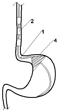
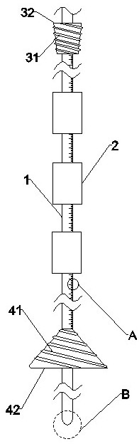
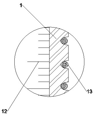
[0034] like figure 1 Shown is a multi-lumen multi-capsule tube capable of fixed-point hemostasis, which is used to solve the problem that the prior art three-chamber two-capsule tube cannot effectively determine the bleeding point, and the hemostatic balloon is large in volume and easily compresses the undamaged part. The structure includes a catheter 1, a hemostatic bag 2, a positioning bag, and a gastric bag 4. The hemostatic bag 2 is located on the outside of the middle part of the catheter 1. After inflating, the hemostatic bag 2 will expand and compress the bleeding site, thereby stopping the bleeding of the esophagus The hemostatic sac 2 is composed of multiple hemostatic sacs 2 with smaller lengths. After the bleeding location is determined through the nasogastroscope 5, only the hemostatic sac 2 close to the bleeding location is used for hemostasis. The compression of the site realizes fixed-point hemostasis, and the patient feels less discomfort. At the same time, the...


PUM


Abstract

The invention discloses a multi-cavity and multi-capsule tube capable of fixed-point hemostasis, which is used to solve the problem that the three-cavity and two-capsule tube in the prior art cannot effectively determine the bleeding point, and the hemostatic air bag has a large volume and is easily compressed into a damaged part. The structure includes a catheter, a hemostatic sac, a positioning sac, and a gastric sac. The hemostatic sac is outside the middle of the catheter. After inflation, the hemostatic sac will expand and compress the bleeding site, thereby stopping the bleeding of the esophagus. The hemostatic sac uses a length of It is composed of multiple smaller hemostatic sacs. After the bleeding location is determined by endoscopy, only the hemostatic sac close to the bleeding location is used for hemostasis. The length of the hemostatic sac is small, which reduces the pressure on undamaged parts and achieves fixed-point hemostasis. The discomfort is less, and at the same time, the positioning bag on the top of the hemostatic bag is inflated and fixed in the esophagus to avoid the movement of the hemostatic bag caused by the downward movement of the catheter. In addition, the contact area between the tapered bag and the stomach is larger, and it is also better for gastric fundus bleeding use effect.

Description

technical field [0001] The invention belongs to the technical field of medical devices, and in particular relates to a multi-cavity multi-capsule tube capable of fixed-point hemostasis. Background technique [0002] For patients with esophageal and gastric fundus variceal rupture caused by cirrhosis and portal hypertension in the acute stage or patients with a large amount of bleeding and unable to maintain effective blood volume, the treatment is mainly to use three-lumen two-capsule catheter for compression hemostasis, which has a significant hemostatic effect and is the first choice The basic treatment mechanism of the three-lumen two-capsule tube is that the gastric tube has an esophageal balloon and a gastric balloon. After inflation, the soft balloon pressure is used to respectively compress the lower esophagus and the varicose veins bleeding from the gastric fundus to achieve hemostasis. Purpose: The length of the esophageal balloon of the three-chamber two-capsule tu...

Claims


Application Information

Patent Timeline
Patent Type & Authority Patents(China)
IPC IPC(8): A61M25/10A61B17/12A61B1/273
CPCA61M25/1011A61B17/12A61B1/2736A61B1/2733A61B2017/12004A61M2210/1053A61M2210/005
Inventor 蒲虹张述蓉赵佑会
Owner WEST CHINA HOSPITAL SICHUAN UNIV
Why Eureka
Try Eureka