Tumor ultrasonic image classification method and device based on optical density transformation, and medium

An ultrasound image and classification method technology, applied in the field of tumor ultrasound image classification, can solve the problems of low foreground discrimination, high gray level, and low image background and foreground discrimination, so as to improve accuracy and robustness, and reduce misdiagnosis. The effect of missed diagnosis and improved diagnosis efficiency

Pending Publication Date: 2020-09-22
HUAQIAO UNIVERSITY +2
View PDF3 Cites 3 Cited by
  • Summary
  • Abstract
  • Description
  • Claims
  • Application Information

AI Technical Summary

Problems solved by technology

The current imaging method of ultrasound images will result in a small distinction between the background and the foreground of the image, high and close gray levels, and it is difficult to distinguish the difference between similar gray areas in the ultrasound image
At present, most studies do not pay attention to the problem of low discrimination between image background and foreground, and only study single-type features or directly fuse two types of features, which tends to ignore important tumor information, resulting in inaccurate automatic classification of tumor ultrasound images

Method used


Image

  • Tumor ultrasonic image classification method and device based on optical density transformation, and medium
  • Tumor ultrasonic image classification method and device based on optical density transformation, and medium
  • Tumor ultrasonic image classification method and device based on optical density transformation, and medium

Examples


Embodiment 1

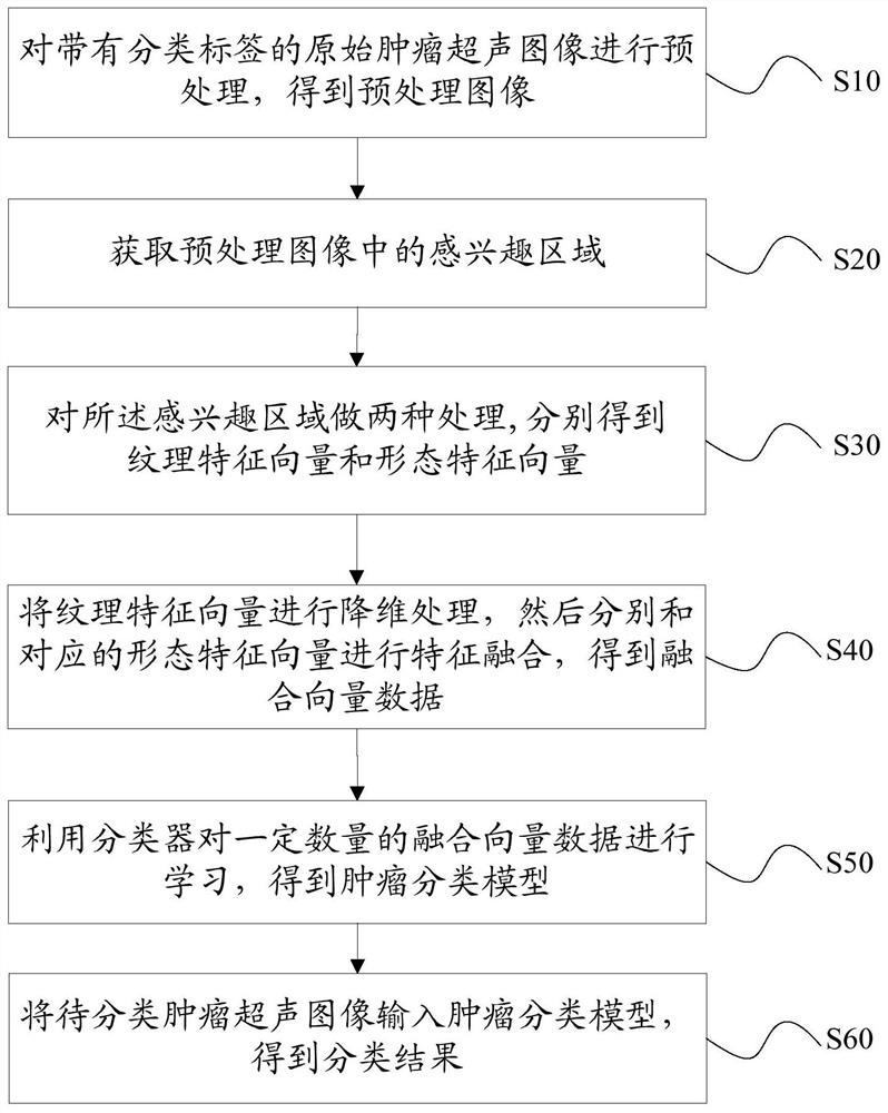
[0058] The present invention provides a tumor ultrasound image classification method based on optical density transformation, such as figure 1 shown, including:

[0059] Step 10, preprocessing the original tumor ultrasound image with classification labels to obtain a preprocessed image;

[0060] In a possible implementation manner, the classification labels include malignant tumors and benign tumors, and the preprocessing includes removing text and personal information around the ultrasound image, reducing speckle noise, and enhancing contrast.

[0061] Step 20, obtaining the region of interest (ROI) in the preprocessed image;

[0062] In a possible implementation manner, a region designated by a doctor in the preprocessed image is acquired as the region of interest.

[0063] By obtaining the region of interest designated by the doctor, the suspicious region can be accurately processed, and the consumption of computing resources caused by invalid data processing is reduced. ...

Embodiment 2

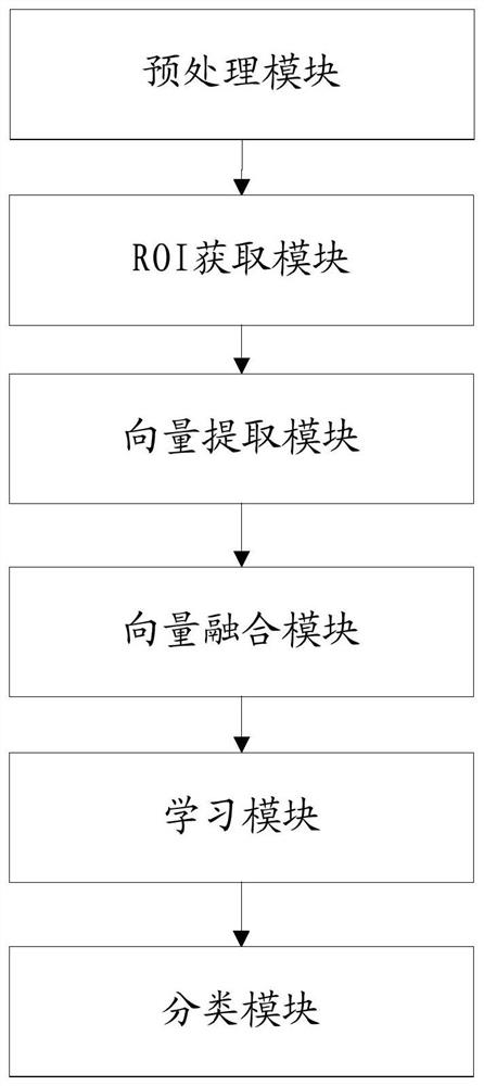
[0090] The present invention provides a tumor ultrasonic image classification device based on optical density transformation, such as figure 2 As shown, including: preprocessing module, ROI acquisition module, vector extraction module, vector fusion module, learning module and classification module;

[0091] The preprocessing module is used to preprocess the original tumor ultrasound images with classification labels to obtain preprocessed images;

[0092] The ROI acquisition module is used to acquire a region of interest (ROI) in the preprocessed image;

[0093] The vector extraction module is used to perform the following two types of processing on the region of interest:

[0094] The first processing: performing optical density transformation on the region of interest to obtain an optical density image, then extracting texture features from the optical density image, and then performing a normalization operation to obtain a texture feature vector;

[0095] The second pro...

Embodiment 3

[0102] The present invention provides a computer-readable storage medium, such as image 3 As shown, a computer program is stored thereon, and when the program is executed by a processor, the method described in the first aspect is realized.


PUM


Abstract

The embodiment of the invention discloses a tumor ultrasonic image classification method and device based on optical density transformation, and a medium, and the method comprises the steps: step 10,carrying out the preprocessing of an original tumor ultrasonic image with a classification label, and obtaining a preprocessed image; step 20, acquiring a region of interest in the preprocessed image;step 30, performing two kinds of processing on the region of interest to obtain a texture feature vector and a morphological feature vector respectively; step 40, performing dimension reduction processing on the texture feature vectors, and performing feature fusion on the texture feature vectors and the corresponding morphological feature vectors to obtain fusion vector data; step 50, learning acertain amount of fusion vector data by using a classifier to obtain a tumor classification model; and step 60, inputting the tumor ultrasonic images to be classified into the tumor classification model to obtain a classification result. The method disclosed by the invention can effectively improve the accuracy and robustness of ultrasonic image tumor benign and malignant prediction, provides reference for doctors to diagnose tumors, and improves the diagnosis efficiency of doctors.

Description

technical field [0001] The present invention relates to the technical field of medical image processing, in particular to a tumor ultrasound image classification method, device and medium based on optical density transformation. Background technique [0002] Cancer is one of the most important diseases threatening human health worldwide. According to the statistics of the World Health Organization, the incidence of cancer has been on the rise. Early detection, early diagnosis and early treatment are the key to improving the cure rate of cancer and reducing the mortality rate. Ultrasound imaging technology is widely used in the detection of tumors because of its versatility, safety and high sensitivity. , Breast Ultrasound, Liver Ultrasound, Heart Ultrasound and Kidney Ultrasound, etc. [0003] Ultrasound is the preferred imaging examination and preoperative evaluation method for the diagnosis of tumors. However, correct interpretation of ultrasound images requires doctors ...

Claims


Application Information

Patent Timeline
IPC IPC(8): G06K9/62G06K9/46
CPCG06V10/44G06V2201/032G06F18/2135G06F18/214G06F18/24G06F18/253
Inventor 杜永兆魏梦婉柳培忠苏淇琛朱建清郭明辉
Owner HUAQIAO UNIVERSITY
Why Eureka
Try Eureka