A kind of ischemic retinopathy detection biomarker, detection kit and application
A technology for retinopathy and ischemia, applied in the field of biomedical detection, which can solve the problems such as the lack of circRNA-Wdr37
- Summary
- Abstract
- Description
- Claims
- Application Information
AI Technical Summary
Problems solved by technology
Method used
Image

Examples
Embodiment 1
[0036] Example 1 Verification of the correlation between circRNA-Wdr37 and ischemic retinopathy
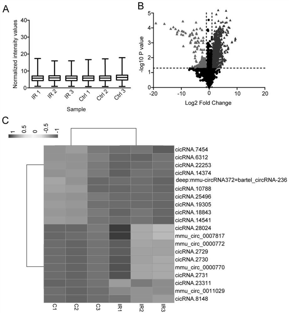
[0037] CircRNA microarray analysis screens and verifies circRNAs related to ischemic retinopathy.
[0038] The first step: sample preparation: the mouse retinal ischemia-reperfusion (IR) animal model was constructed, and the retinal tissues of the experimental group (IR, n=3) and the control group (Ctrl, n=3) were collected. RNA was extracted with TRIzol (Invitrogen) reagent and stored at -80°C for future use.
[0039] Step 2: Differentially expressed circRNA screening:
[0040] Using the circRNA expression profile chip of Aglient Company in the United States, the circRNA related to the occurrence of ischemic retinopathy was analyzed; the specific steps of the analysis were: using a labeling enzyme to label the circRNA with a fluorescent group to obtain a fluorescent probe for hybridization with the chip. Use the MAUI hybridization instrument and chip hybridization; use the Gene...
Embodiment 2
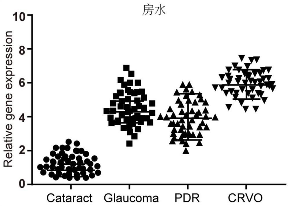
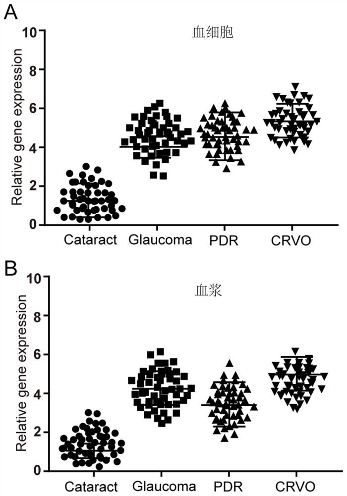
[0045] Example 2 Detection of expression of circRNA-Wdr37 in blood cells and plasma
[0046] Step 1: Separation of serum and plasma samples
[0047] Blood samples of 200 cases of each patient in the experimental group (primary open-angle glaucoma, central vein occlusion, and diabetic retinopathy) and the control group (cataract) were collected, and serum and serum were separated by centrifugation using heparin anticoagulation tubes. Plasma RNA for detection of circRNA. The centrifugation conditions were 4°C, 12,000 rpm, 10 min.
[0048] Step 2: RNA Extraction from Serum and Plasma Samples
[0049] Add TRIzol to the separated blood cells and plasma samples respectively, and place at room temperature for 10 minutes to fully lyse the samples (Note: If the next step is not performed, the samples can be stored at -70°C for long-term storage). Add 200 μl of chloroform to every 1ml of TRIzol, vibrate vigorously and mix well, then place at room temperature for 3-5min to allow natur...
Embodiment 3
[0075] Example 3 Feasible application of circRNA-Wdr37 as a marker for prognosis assessment
[0076] The first step; obtain the blood sample to be tested
[0077] Sera from 200 patients with each disease were collected before and after treatment for ischemic retinopathy (primary open-angle glaucoma, central vein occlusion, and diabetic retinopathy).
[0078] Step 2: Extract RNA from the blood sample to be tested
[0079] a) Serum sample or frozen serum Take 0.25ml sample and transfer it to a centrifuge tube, add 1ml Trizol, pipette up and down repeatedly until the cells are completely lysed.
[0080] b) Add chloroform (sample solution + 1 / 5 volume of Trizol Reagent volume) to cover the centrifuge tube tightly, vibrate vigorously for 15 sec, and let stand at room temperature for 5 min;
[0081] c) Centrifuge at 4°C, 12,000g×15min. Carefully take out the centrifuge tube from the centrifuge. At this time, the homogenate is divided into three layers, namely: a colorless supernat...
PUM
| Property | Measurement | Unit |
|---|---|---|
| Sensitivity | aaaaa | aaaaa |
Abstract
Description
Claims
Application Information
 - R&D Engineer
- R&D Manager
- IP Professional
- Industry Leading Data Capabilities
- Powerful AI technology
- Patent DNA Extraction
Browse by: Latest US Patents, China's latest patents, Technical Efficacy Thesaurus, Application Domain, Technology Topic.
© 2024 PatSnap. All rights reserved.Legal|Privacy policy|Modern Slavery Act Transparency Statement|Sitemap