Oral cavity examination device for department of oral medicine

An inspection device and oral medicine technology, applied in the field of medical devices, can solve the problems of unable to support the mouth, inconvenient for doctors to observe and use, and blocking the doctor's line of sight, etc.

Inactive Publication Date: 2020-11-20
HENAN MEDICAL COLLEGE
View PDF10 Cites 3 Cited by
  • Summary
  • Abstract
  • Description
  • Claims
  • Application Information

AI Technical Summary

Problems solved by technology

Existing intraoral endoscopes have a handle. During the inspection, the patient needs to open his mouth and extend the oral endoscope into the oral cavity for inspection. This inspection method cannot support t

Method used


Image

  • Oral cavity examination device for department of oral medicine
  • Oral cavity examination device for department of oral medicine
  • Oral cavity examination device for department of oral medicine

Examples


Embodiment 1

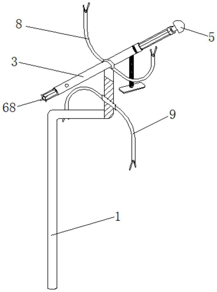
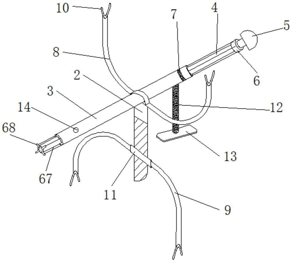
[0037] The invention provides an oral examination device for oral medicine, specifically as Figure 1 to Figure 6 As shown, including support rod 1, inspection module and support module;

[0038]The upper end of the support rod 1 is provided with a section of external thread, and the lower end is an L-shaped structural member arranged in a horizontal mirror image;

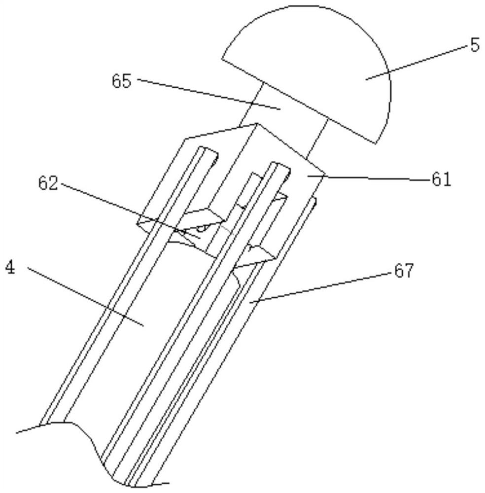
[0039] The inspection module includes a connecting column 2, an outer sleeve 3, an inner sleeve 4, an oral endoscope 5 and an angle adjustment mechanism 6; the lower end of the connecting column 2 is rotationally connected with the upper end of the support rod 1, and the upper end is movably connected with the outer sleeve 3; the inner sleeve 4 is set in the outer sleeve 3 and is slidingly connected with the outer sleeve 3, and both ends extend out from the two ends of the outer sleeve 3. The oral endoscope 5 is connected to one end of the inner sleeve 4 through an angle adjustment mechanism 6, and is far away from...

Embodiment 2

[0052] To facilitate device placement, such as Figure 7 As shown, in this embodiment, the lower end of the support rod 1 is sleeved with an L rod 14 telescopically connected to the support rod 1, and a plurality of first position-limiting holes 15 are provided on the longitudinal rod of the L rod 14, and the support rod 1 and the vertical The rod is fixed by the first limit bolt 16, and the end of the transverse rod of the L rod 14 is provided with a fixing clip 17, and the whole device is clamped on the edge of the table through the fixing clip 17, and the hand-holding device is no longer used, and the rest of the structure is the same as in Example 1. Same, no more details here.

Embodiment 3

[0054] To facilitate device placement, such as Figure 8 As shown, in this embodiment, the lower end of the support rod 1 is sleeved with a vertical rod 18 that is telescopically connected to the support rod 1, and the support rod 1 is provided with a plurality of second limit holes 19, and the support rod 1 and the vertical rod pass through the first Two limit bolts 20 are fixed, and the lower end of the vertical rod 18 is provided with a triangular support frame 21 with self-locking rollers. The whole device can be moved conveniently by the tripod 21, and the whole device is placed on the desktop and other positions by the tripod 21, and the hand-held device is no longer used. The rest of the structure is the same as in Embodiment 1, and will not be repeated here.


PUM


Abstract

The invention provides an oral cavity examination device for the department of oral medicine, and belongs to the technical field of medical apparatus and instruments. The lower end of a connecting column is rotationally connected with the upper end of a supporting rod, and the upper end of the connecting column is movably connected with an outer sleeve; an inner sleeve is sleeved with the outer sleeve, an oral cavity endoscope is connected with one end of the inner sleeve through an angle adjusting mechanism and is away from an L-shaped structural rod piece; a ring-shaped LED lamp is arrangedat the end, close to the oral cavity endoscope, of the outer sleeve in the axial direction, and a tongue press mechanism is arranged at the bottom of the outer sleeve and close to the LED lamp; a supporting module comprises an upper supporting body and a lower supporting body which have the same structure and arranged in a vertically symmetric manner, the upper supporting body is of a U-shaped structure, and V-shaped clamp grooves are formed in the tops of two ends of the upper supporting body; the middle of the upper supporting body is located above a groove and fixedly connected with a supporting rod; and a slider is in screw joint with the upper end of the supporting rod, and the middle of the lower supporting body is fixedly connected with the slider. According to the oral cavity examination device for the department of oral medicine, pathologic conditions in the oral cavity can be collected in a multidirectional manner by the oral cavity endoscope and the angle adjusting mechanism, meanwhile, the sight of a doctor cannot be blocked, and observation and use of the doctor are convenient.

Description

technical field [0001] The invention belongs to the technical field of medical devices, and in particular relates to an oral examination device for oral medicine. Background technique [0002] As one of the most important structures in the human body, the oral cavity is the beginning part of the digestive tract, consisting of two lips, two cheeks, hard palate, and soft palate. It communicates with the outside world through the cleft of the mouth, and connects with the pharynx through the pharynx. There are teeth, tongue and other organs in the oral cavity. Since the oral cavity is connected to the outside world, it is easily infected by external bacteria. It is a place where the human body is more prone to diseases, resulting in various diseases such as periodontal and oral mucosa. Therefore, it is very necessary to check the inside of the oral cavity regularly. [0003] A common oral examination is that the doctor uses a tongue depressor to depress the tongue, takes a pict...

Claims


Application Information

Patent Timeline
IPC IPC(8): A61B1/24A61B1/06A61B1/04
CPCA61B1/0684A61B1/04A61B1/24A61B1/00154
Inventor 王凯亭李建斌
Owner HENAN MEDICAL COLLEGE
Why Eureka
Try Eureka