Application of nuclear export protein inhibitor kpt-335 and its composition in antitumor drugs
A KPT-335, protein inhibitor technology, applied in the field of biomedicine to achieve good inhibitory effect, improved curative effect, and low toxicity
- Summary
- Abstract
- Description
- Claims
- Application Information
AI Technical Summary
Problems solved by technology
Method used
Image
Examples
Embodiment 1
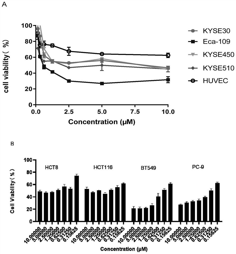
[0025] Embodiment 1, the effect of KPT-335 single use on the proliferation of tumor cells and normal cells
[0026] Esophageal cancer cell lines KYSE30, Eca-109, KYSE450, KYSE510, breast cancer cell line BT549, lung cancer cell line PC-9, colorectal cancer cell lines HCT8, HCT116 and normal vascular endothelial cells HUVEC were used at 3000-6000 per well The number of cells was inoculated into a 96-well plate. After the cells adhered to the wall, the blank control group and different doses of KPT-335 experimental groups (0-10 μM) were added; after 48 hours of culture, 10 μL of CCK-8 solution was added to each well, and the culture was continued for 1 hour , read the OD value (wavelength 450nm) on a microplate reader, read the absorbance value of each well, and calculate the cell viability. According to the formula of cell survival rate (%)=(OD experimental group-OD blank) / (OD control group-OD blank)×100%, the cell survival rate is calculated, and the results are detailed in f...
Embodiment 2
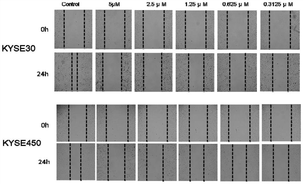
[0030] Embodiment 2, the effect of KPT-335 alone on the migration of esophageal cancer cells
[0031] Esophageal cancer cell lines KYSE30 and KYSE450 were inoculated into 96-well plates at the number of 12,000 cells per well. After the cells adhered to the wall, they were synchronized for 12 hours, scratched with a scratch instrument, washed twice with PBS, and replaced with 2% Fetal bovine serum medium, take pictures, this time is 0h, then add the blank control group and the KPT-335 experimental group with doses of 0, 5, 2.5, 1.25, 0.625, 0.31125 μM respectively, take pictures after 24 hours of culture, measure Width, calculate mobility. According to cell migration ratio=width (experimental group 0h-experimental group 24h) / width (blank group 0h-blank group 24h) × 100%, the results are shown in Table 2 and figure 2 .
[0032] Table 2 The results of each cell migration rate measurement
[0033]
[0034]
[0035] From Table 2 and figure 2It can be seen that 5 μM KPT-...
Embodiment 3
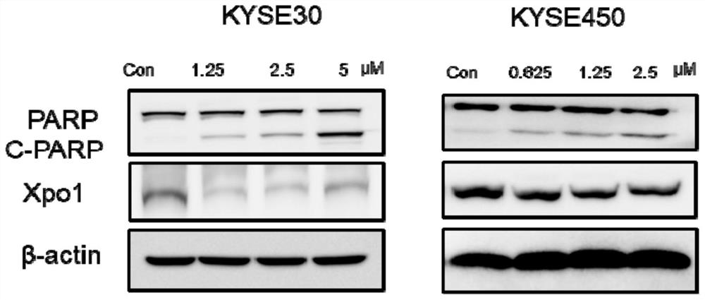
[0036] Embodiment 3, the effect of KPT-335 alone on the apoptosis protein of esophageal cancer cells
[0037] First, the esophageal cancer cells KYSE30 and KYSE450 were divided into 3.5×10 5 cells at a density of 6 wells. After the cells adhered to the wall, they were treated with different concentrations for 24 hours, and the total protein of the cells in each group was collected respectively. After SDS electrophoresis, the protein was transferred to a PVDF membrane, blocked with 5% skimmed milk powder at room temperature for 1 hour, incubated with the primary antibody corresponding to the protein to be detected, and incubated overnight at 4°C. After 24 hours, the primary antibody was recovered, washed three times with TBST for 5 minutes each time, and then incubated with the secondary antibody for 1 hour at room temperature. After washing with TBST for 3 times for 5 min each time, ECL imaging was performed to detect the expression of apoptosis pathway-related proteins PARP...
PUM
 Abstract
Description
Claims
Application Information
 - R&D
- Intellectual Property
- Life Sciences
- Materials
- Tech Scout
- Unparalleled Data Quality
- Higher Quality Content
- 60% Fewer Hallucinations
Browse by: Latest US Patents, China's latest patents, Technical Efficacy Thesaurus, Application Domain, Technology Topic, Popular Technical Reports.
© 2025 PatSnap. All rights reserved.Legal|Privacy policy|Modern Slavery Act Transparency Statement|Sitemap|About US| Contact US: help@patsnap.com


