Automatic delineation method for organs at risk in pelvic radiotherapy
An organ and pelvic technology, applied in the field of automatic delineation of organs at risk in pelvic radiotherapy, can solve the problems of low efficiency of manual delineation, poor repeatability, and dependence on experience level, etc., to improve prediction accuracy and generalization ability, improve consistency, and improve The effect of precision
- Summary
- Abstract
- Description
- Claims
- Application Information
AI Technical Summary
Problems solved by technology
Method used
Image
Examples
Embodiment 1
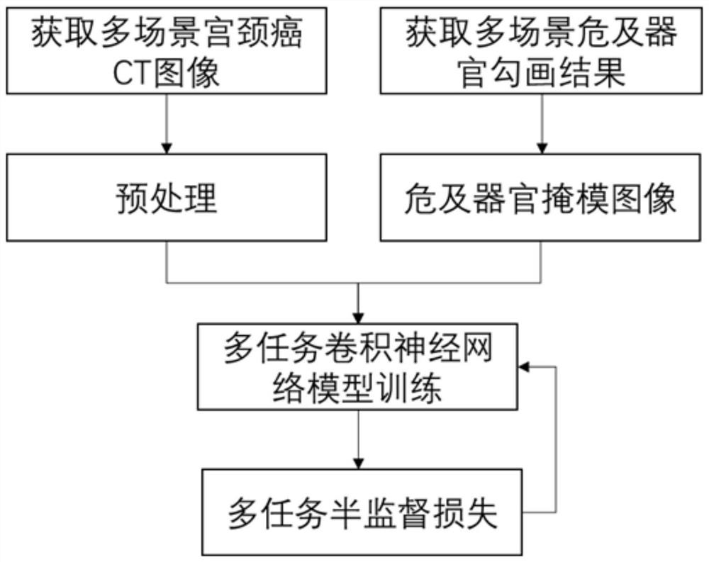
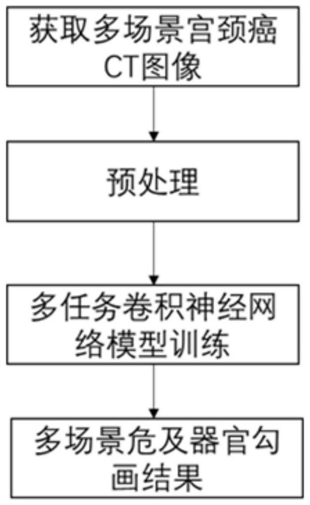
[0037] This example provides a method for automatic delineation of organs at risk in pelvic radiotherapy, including the following steps:
[0038] S1: Obtain the CT image data of the external irradiation plan and the CT image data of the intracavitary irradiation plan of the cervical cancer patients in the training data set, and perform preprocessing on the CT image data of the external irradiation plan and the CT image data of the intracavitary irradiation plan;
[0039] S2: Extract the outline of each organ at risk outlined on the CT image of the external irradiation plan of cervical cancer patients in the training data set, and assign the area inside the outline as 1 and the area outside the outline as 0, and obtain the binary value of the organ at risk in external irradiation mask image;
[0040] S3: Extract the contour of each organ at risk outlined on the CT image of the endocavitary irradiation plan of cervical cancer patients in the training data set, and assign the are...
Embodiment 2
[0063] Based on Example 1, this embodiment compares the present invention: a method for automatically delineating organs at risk in pelvic radiotherapy with other conventional methods, including the following steps:
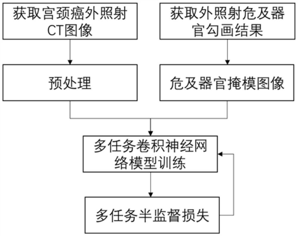
[0064] S1: The external radiation plan CT image data of cervical cancer patients and the binary mask image of organs at risk in the training data set obtained and processed in steps S1-S4 in Example 1 are performed on the convolutional neural network with the same structure as the present invention. training, the training process is as follows image 3 As shown, the delineation model A of organs at risk for external exposure is obtained;
[0065] S2: The intracavity irradiation plan CT image data of cervical cancer patients and the binary mask image of organs at risk obtained and processed in the training data set obtained and processed in steps S1-S4 in Example 1 are paired with the convolutional neural network with the same structure as the present invention F...
PUM
 Abstract
Description
Claims
Application Information
 - R&D
- Intellectual Property
- Life Sciences
- Materials
- Tech Scout
- Unparalleled Data Quality
- Higher Quality Content
- 60% Fewer Hallucinations
Browse by: Latest US Patents, China's latest patents, Technical Efficacy Thesaurus, Application Domain, Technology Topic, Popular Technical Reports.
© 2025 PatSnap. All rights reserved.Legal|Privacy policy|Modern Slavery Act Transparency Statement|Sitemap|About US| Contact US: help@patsnap.com


