Intracranial hemorrhage automatic sketching method and device based on head CT (Computed Tomography) plain-scan image
A technology for intracranial hemorrhage and head, which is applied in the field of AI medical image-aided diagnosis, can solve the problems such as subarachnoid hemorrhage segmentation is difficult to reflect, bleeding segmentation performance is not ideal, and complete segmentation is difficult, so as to ensure the feasibility of optimization and reduce diagnosis The time required to improve the effect of accurate diagnosis
- Summary
- Abstract
- Description
- Claims
- Application Information
AI Technical Summary
Problems solved by technology
Method used
Image
Examples
Embodiment 1
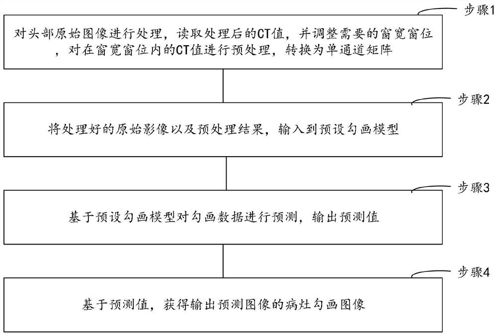
[0100] The present invention provides a method for automatic delineation of intracranial hemorrhage based on head CT plain scan images, such as figure 1 shown, including:
[0101] Step 1: Process the original image of the head, read the processed CT value, adjust the required window width and level, preprocess the CT value within the window width and level, and convert it into a single-channel matrix;
[0102] Step 2: Input the processed original image and preprocessing results into the preset sketching model;
[0103] Step 3: Predict the sketched data based on the preset sketching model, and output the predicted value;
[0104] Step 4: Based on the prediction value, obtain a lesion delineation image of the output prediction image.
[0105] In this example, when the doctor diagnoses intracranial hemorrhage, the main window width and window level used can be the brain window [40,80], and the subdural window level will be used when diagnosing subdural hemorrhage, which can be ...
Embodiment 2
[0111] On the basis of Embodiment 1, step 1: process the original image of the head, read the processed CT value, and adjust the required window width and level, and preprocess the CT value within the window width and window level, Convert to a single-channel matrix, including:
[0112] Read the CT value of the original image of the head, filter the value that meets the first condition in the CT value and keep it, and remove the remaining value to obtain the coordinate information of the skull;
[0113] Set the required window width and window level, and then extract the first value from the CT value of the original head image;
[0114] The extracted first value is normalized, and the coordinate information of the skull is masked in the matrix corresponding to the set window width and level, and converted to 0 to construct a single-channel matrix.
[0115] In this embodiment, normalization is performed on the extracted value to convert the value between 0 and 1, and the previ...
Embodiment 3
[0121] On the basis of Embodiment 1, in step 2, before inputting into the preset sketching model, it also includes:
[0122] Construct the basic intracranial hemorrhage segmentation model and determine the segmentation network evaluation index set;
[0123] Based on the evaluation index set, evaluate the basic intracranial hemorrhage segmentation model;
[0124] According to the evaluation results, determine the basic level of the basic intracranial hemorrhage segmentation model;
[0125] When the basic level is the normal level, the basic intracranial hemorrhage segmentation model is optimized to obtain a preset sketching model;
[0126] When the basic level is a special level, the basic intracranial hemorrhage segmentation model is saved, and the saved model is the preset sketching model.
[0127] In this embodiment, the construction of the basic intracranial hemorrhage segmentation model, such as image 3 As shown, it can be achieved as follows:
[0128] The convolution...
PUM
 Abstract
Description
Claims
Application Information
 - R&D
- Intellectual Property
- Life Sciences
- Materials
- Tech Scout
- Unparalleled Data Quality
- Higher Quality Content
- 60% Fewer Hallucinations
Browse by: Latest US Patents, China's latest patents, Technical Efficacy Thesaurus, Application Domain, Technology Topic, Popular Technical Reports.
© 2025 PatSnap. All rights reserved.Legal|Privacy policy|Modern Slavery Act Transparency Statement|Sitemap|About US| Contact US: help@patsnap.com


