
Convenient-to-use film viewer for oncology department clinical examination

A technology of clinical examination and film viewer, which is applied in the direction of optical components, optics, instruments, etc., can solve the problems of difficult to distinguish, high diagnosis error rate, etc., and achieve the effect of reducing the error rate

Inactive Publication Date: 2020-12-25
刘晓
View PDF9 Cites 0 Cited by
  • Summary
  • Abstract
  • Description
  • Claims
  • Application Information

AI Technical Summary

Problems solved by technology

[0002] When an oncologist looks at the negatives, it seems easy to distinguish between normal and pathological, but it is actually not easy, and sometimes it is very difficult, because the range of normal and normal variation is extremely wide, even those who have worked for decades still need to be vigilant at all times Strictly distinguish between the two, many of these cases looked like lesions yesterday, but they are actually within the normal range. Some bone infections are very similar to the X-ray manifestations of early malignant tumors, and it is difficult to distinguish them. The existing way for doctors to view negative films is Clamp the negative film under the viewing light to observe the negative film, and when observing, the doctor only relies on observation to make a diagnosis, and the diagnosis error rate is high

Method used


Image

  • Convenient-to-use film viewer for oncology department clinical examination
  • Convenient-to-use film viewer for oncology department clinical examination
  • Convenient-to-use film viewer for oncology department clinical examination

Examples


Embodiment

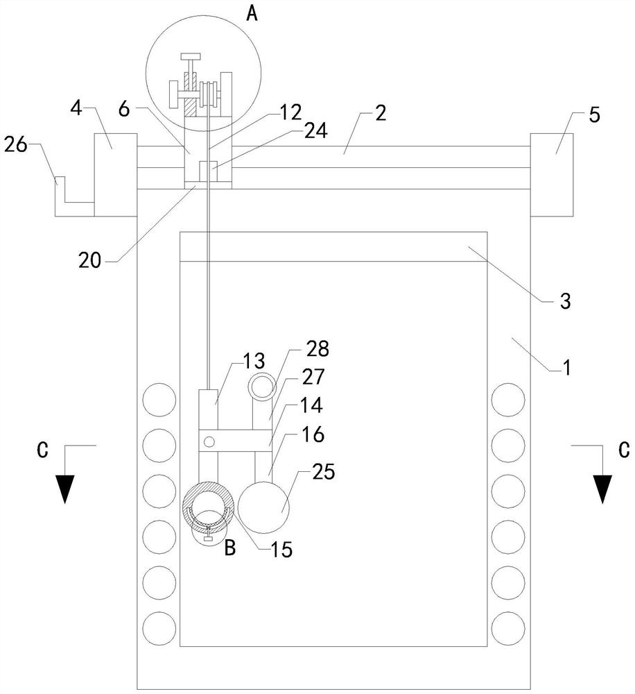
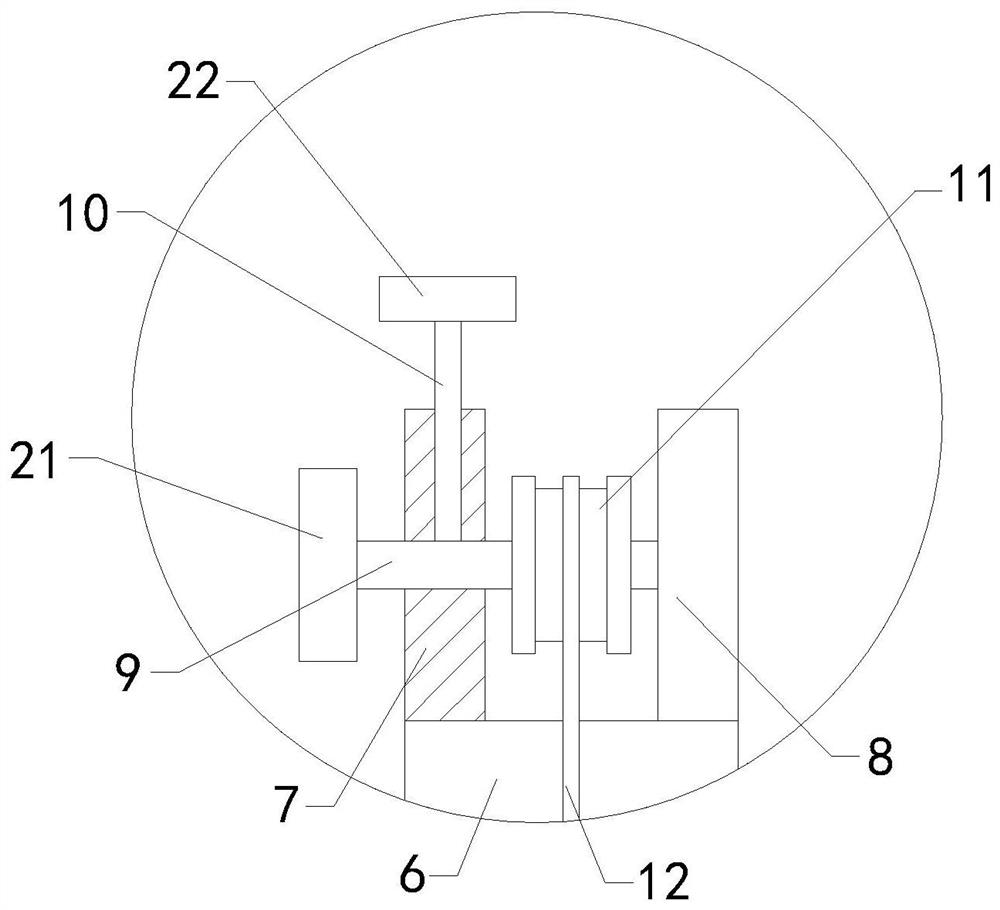
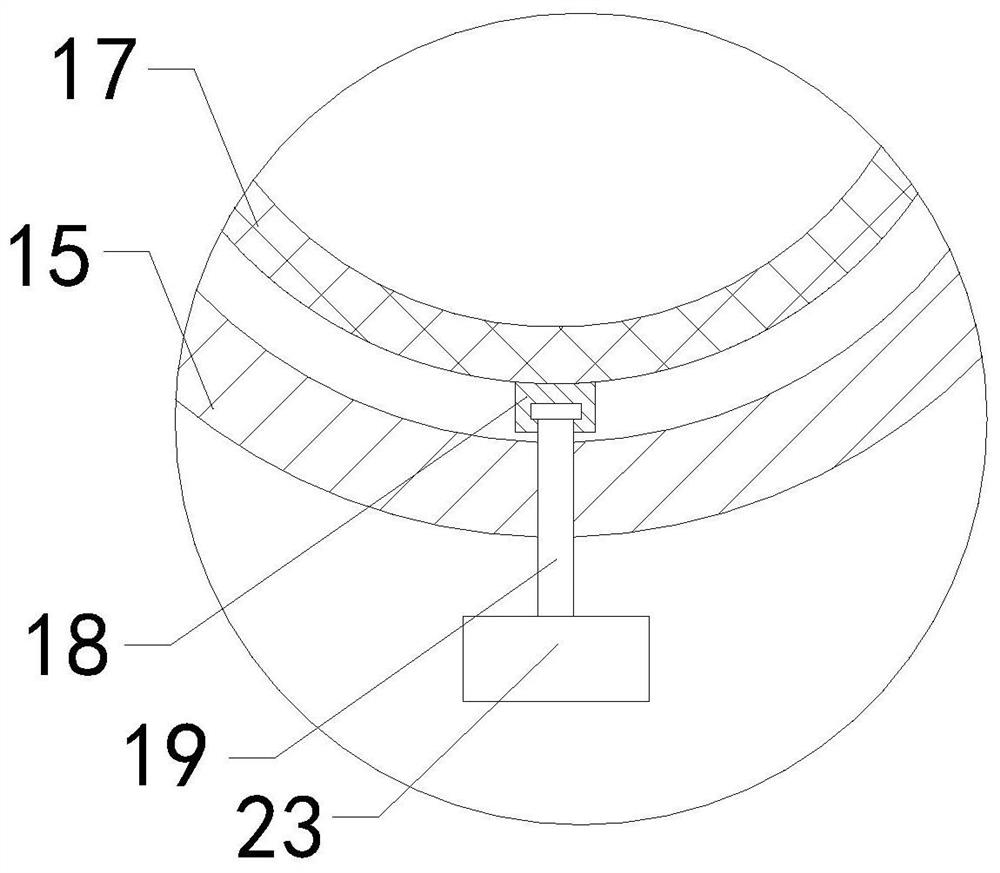
[0025]SeeFigure 1-5, An easy-to-use film viewer for clinical examinations in the department of oncology, comprising a frame body 1 and a limiting shaft 2. A groove is provided in the middle of the frame body 1, a lamp tube 3 is installed on the top of the inner wall of the frame body 1, and the frame body 1 The left and right parts of the front end of the frame body 1 are both provided with installation grooves, the upper left end of the frame body 1 is fixedly connected with a first support plate 4, the upper right end of the frame body 1 is fixedly connected with a second support plate 5, and the left end of the limit shaft 2 It is fixedly connected to the right end of the first support plate 4, and the right end of the limit shaft 2 is fixedly connected to the left end of the second support plate 5. The outer circumferential wall of the limit shaft 2 is slidably connected to the sliding block 6 and the bottom end of the sliding block 6 is fixed. A wear pad 20 is connected. The we...


PUM


Abstract

The invention relates to the technical field of oncology department film viewers, in particular to a convenient-to-use film viewer for oncology department clinical examination. The convenient-to-use film viewer comprises a frame body and a limiting shaft, a groove is formed in the middle of the frame body, a lamp tube is mounted at the top end of the inner wall of the frame body, mounting groovesare correspondingly formed in the left part and the right part of the front end of the frame body, a first supporting plate is fixedly connected to the upper portion of the left end of the frame body,a second supporting plate is fixedly connected to the upper portion of the right end of the frame body, the left end of the limiting shaft is fixedly connected with the right end of the first supporting plate, the right end of the limiting shaft is fixedly connected with the left end of the second supporting plate, a sliding block is connected to the outer wall of the periphery of the limiting shaft in a left-right sliding mode, the left portion of the top end of the sliding block is fixedly connected with a left frame plate, the right portion of the top end of the sliding block is fixedly connected with a right frame plate, the middle of the left end of the right frame plate is rotationally connected with a rotating shaft, the left end of the rotating shaft penetrates through the middleof the left frame plate from the right end of the left frame plate and extends out of the left side of the left frame plate, and a screw rod is arranged on the upper portion of the left frame plate. According to the convenient-to-use film viewer, the error rate of diagnosis is reduced.

Description

Technical field[0001]The invention relates to the technical field of oncology film viewers, in particular to a convenient film viewer for oncology clinical examinations.Background technique[0002]When oncologists observe the film, it seems easy to distinguish between normal and pathological, but it is not simple. Sometimes it is very difficult. Because the range of normal and normal variation is extremely wide, even those who have worked for decades still need to be vigilant. There is a strict distinction between the two. Many of the cases looked like lesions yesterday, but they are all within a normal range. Some bone infections are very similar to the X-ray manifestations of early malignant tumors and are difficult to distinguish. The current way doctors look at the film is Clamp the film under the viewing lamp to observe the film. During the observation, the doctor only makes diagnosis by observation, and the error rate of diagnosis is high.Summary of the invention[0003](1) Techni...

Claims


Application Information

Patent Timeline
Patent Type & Authority Applications(China)
IPC IPC(8): G02B27/02
CPCG02B27/024G02B27/028
Inventor 刘晓
Owner 刘晓
Learn More