Eureka AIR delivers breakthrough ideas for toughest innovation challenges, trusted by R&D personnel around the world.

A deep learning-based segmentation method for breast cancer pathological images he cancer nest

A pathological image, deep learning technology, applied in the field of deep learning, can solve problems such as time-consuming and discrepancies, and achieve the effect of improving performance, improving efficiency, and enriching semantic information

Active Publication Date: 2022-08-02
WEST CHINA HOSPITAL SICHUAN UNIV +1
View PDF0 Cites 1 Cited by
  • Summary
  • Abstract
  • Description
  • Claims
  • Application Information

AI Technical Summary

Problems solved by technology

In common image reading scenarios, pathologists need to manually find cancer nest areas in a large number of tissue areas based on their own experience, which is time-consuming and varies among different pathologists

Method used


Image

  • A deep learning-based segmentation method for breast cancer pathological images he cancer nest
  • A deep learning-based segmentation method for breast cancer pathological images he cancer nest
  • A deep learning-based segmentation method for breast cancer pathological images he cancer nest

Examples


Embodiment Construction

[0031] In order to make the objectives, technical solutions and advantages of the present invention clearer, the present invention will be further described in detail below with reference to the accompanying drawings.

[0032] The HE WSI described in this paper, the Whole Slide Image, is a fully digitized HE pathological slice image.

[0033] like Figure 8 As shown, the present invention comprises the following steps:

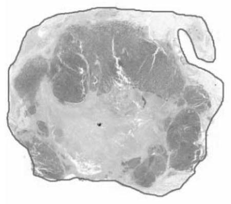
[0034] 1. If figure 1 As shown, an FCN (Fully Convolutional Networks) segmentation network is trained to extract the outline of the effective tissue area in the 1x image, map it to the 40x image, and extract the effective tissue area correspondingly.

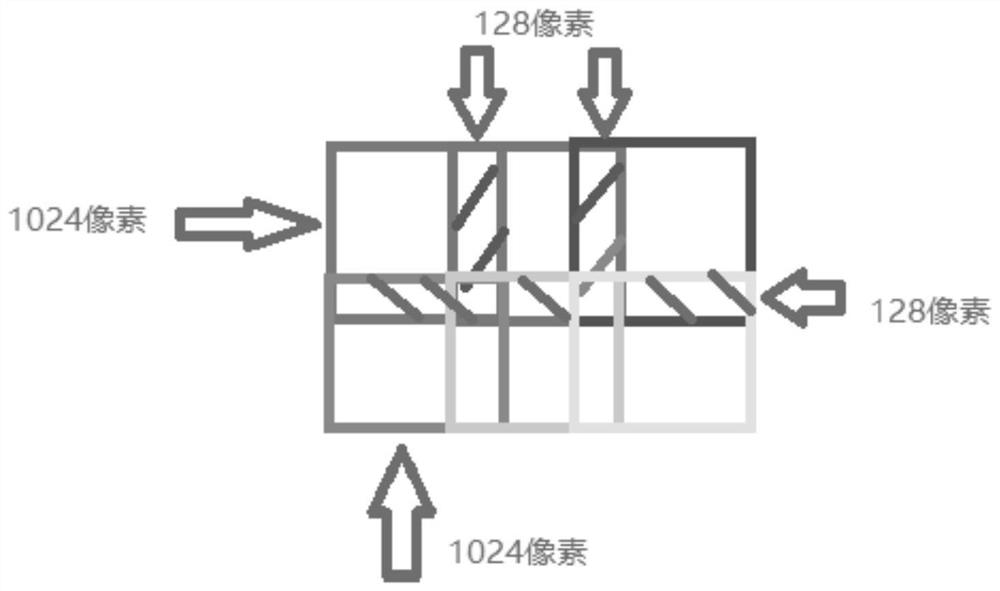
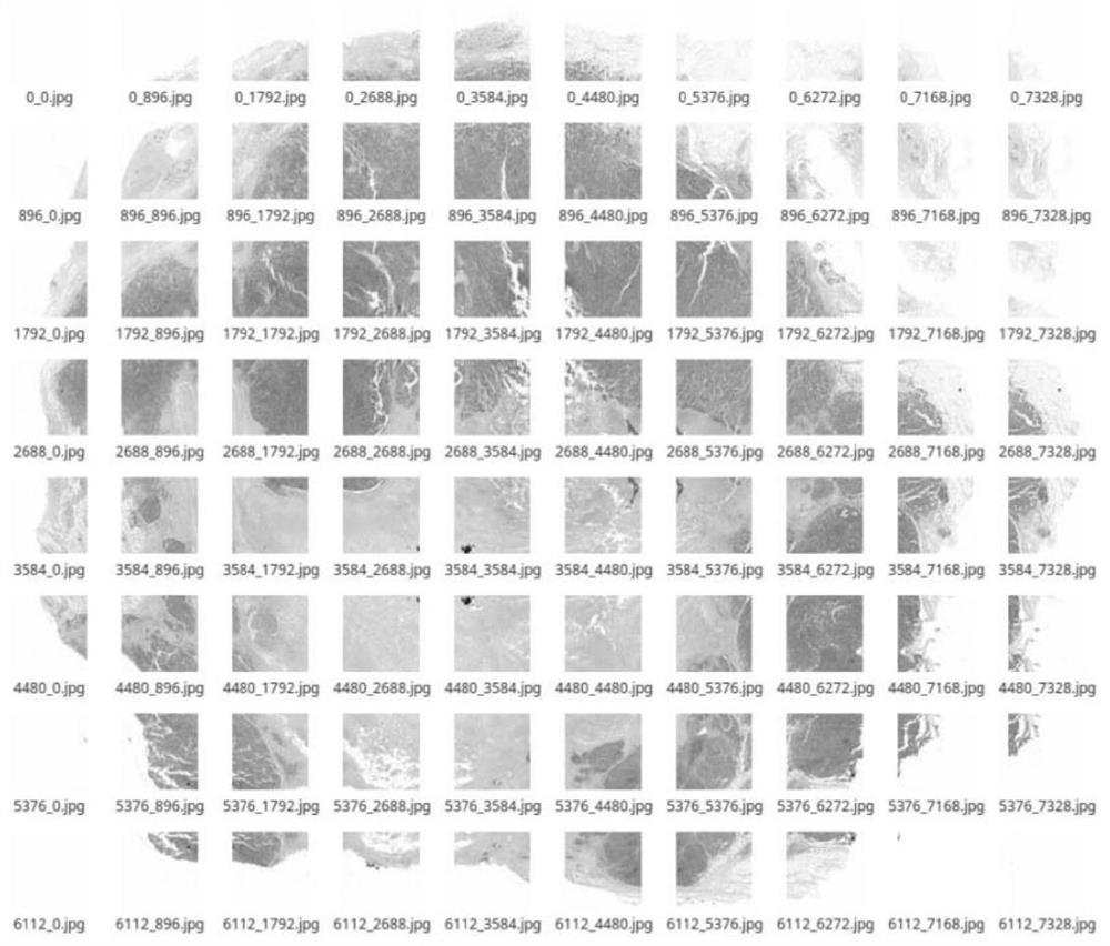
[0035] 2. If figure 2 , image 3 As shown in the figure, the extracted tissue area at a magnification of 40 is oversampled in a manner of overlapping 128 pixels, and the image is cropped into several patches of 1024*1024 (length*width).

[0036] 3. If Figure 5 As shown, the cropped patch at a magnificati...


PUM


Abstract

The invention discloses a deep learning-based method for segmenting a breast cancer pathological image HE cancer nest, comprising: S1, inputting a piece of HE WSI, systematically segmenting a model, and under 1x, segmenting the contour area of ​​the tissue in the slice; S2, segmenting 1x map the area under 40x, and extract the corresponding area; S3, crop the extracted area into a Patch with a size of 1024*1024 and overlap 128 pixels; S4, increase the magnification of all Patches to 80x; S5, increase the height of The resolution results are input into the semantic segmentation model, and the model outputs the segmentation Mask of each Patch; S6, merge each Mask according to the cropped coordinates to generate a complete binary Mask image; S7, perform morphological operations on the merged binary image , and extract contours hierarchically. The invention adopts the deep neural network for segmentation, which has stronger generalization ability and higher robustness, adopts the overlapping method, designs the processing mechanism of the boundary effect, and can effectively avoid the boundary effect.

Description

technical field [0001] The invention relates to the field of deep learning, in particular to a deep learning-based method for segmenting a breast cancer pathological image HE cancer nest. Background technique [0002] In the daily diagnosis of breast cancer, pathologists usually perform HE immunohistochemical slide analysis. Through a comprehensive analysis of the distribution and types of cancer nests in HE images under a microscope, and with other immunohistochemical indicators, a final diagnosis report is given. With the development of digital pathology, HE slices can be scanned into digital pathology slices by a digital scanner. Pathologists can browse pathological slices on the computer through open source or specific image reading tools. The current scanning imaging images are clear, truly reflect the information of slices, and can be stored for a long time. In the process of reading the image, the slice contains a lot of slice tissue information, among which the ca...

Claims


Application Information

Patent Timeline
Patent Type & Authority Patents(China)
IPC IPC(8): G06T7/11G06T7/187G06T7/136G06T3/40G06N3/04G06N3/08
CPCG06T7/11G06T7/187G06T7/136G06T3/4053G06T3/4023G06T3/4038G06N3/08G06T2207/10061G06T2207/20132G06T2207/20021G06N3/045
Inventor 向旭辉郑众喜卫亚妮陈杰王杰步宏
Owner WEST CHINA HOSPITAL SICHUAN UNIV
Why Eureka
Eureka Blog
Learn More