Method and equipment for identifying orientation of intracerebral fragmentation electrode in craniocerebral medical image

A technology of medical imaging and recognition methods, applied in the field of medical imaging, can solve the problems of low judgment accuracy, time-consuming, inconvenient remote consultation, etc., and achieve the effect of saving judgment time, facilitating remote consultation, and improving judgment accuracy

Pending Publication Date: 2021-04-16
SCENERAY
View PDF4 Cites 3 Cited by
  • Summary
  • Abstract
  • Description
  • Claims
  • Application Information

AI Technical Summary

Problems solved by technology

However, to judge the position and direction angle of the sliced ​​electrodes in the brain now, the patient must bring the image film when seeing a doctor. The doctor needs to directly compare the image film to judge the position of the electrode in the brain. The judgment needs to be based on the doctor’s experience. Judgment accuracy is low and time-consuming, and it is not convenient for remote consultation

Method used


Image

  • Method and equipment for identifying orientation of intracerebral fragmentation electrode in craniocerebral medical image

Examples


Embodiment Construction

[0021] The present invention will be further described below in conjunction with the accompanying drawings and specific embodiments, so that those skilled in the art can better understand the present invention and implement it, but the examples given are not intended to limit the present invention.

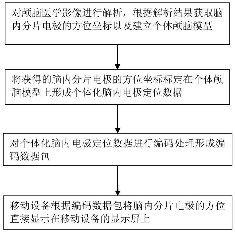
[0022] Such as figure 1 As shown, the present embodiment discloses a method for identifying the orientation of sliced ​​electrodes in the brain in craniocerebral medical imaging, which includes the following steps:

[0023] 1) Analyze the brain medical images, obtain the azimuth coordinates of the slice electrodes in the brain according to the analysis results, and establish an individual brain model (three-dimensional model) according to the analysis results;

[0024] 2) Marking the azimuth coordinates of the obtained intracerebral slice electrodes on the individual brain model to form individualized intracerebral electrode positioning data;

[0025] 3) Coding and processing the...


PUM


Abstract

The invention discloses a method and equipment for identifying the orientation of an intracerebral fragmentation electrode in a craniocerebral medical image. The method comprises the following steps: firstly analyzing the craniocerebral medical image, obtaining the orientation coordinates of the intracerebral fragmentation electrode according to an analysis result, and building an individual craniocerebral model according to the analysis result; calibrating the obtained azimuth coordinates of the intracerebral fragmentation electrodes on an individual craniocerebral model to form individualized intracerebral electrode positioning data; and encoding the individualized intracerebral electrode positioning data to form an encoded data packet, and directly displaying the orientation of the intracerebral fragmentation electrode on a display screen of mobile equipment by the mobile equipment according to the encoded data packet. The invention further discloses the equipment corresponding to the method. According to the invention, the spatial orientation of the intracerebral fragmentation electrode in the brain can be visually displayed on the mobile device, a doctor does not need to compare an image to carry out manual judgment, the judgment accuracy is improved, and the manual judgment time is shortened; and remote inquiry can be carried out conveniently.

Description

technical field [0001] The invention relates to the technical field of medical imaging, in particular to a method and equipment for identifying the orientation of sliced ​​electrodes in the brain in craniocerebral medical imaging. Background technique [0002] In the medical industry, doctors usually use medical images to observe and diagnose patients' symptoms, and brain medical images are an important basis for doctors to judge brain diseases. When doctors observe craniocerebral medical images, they need to measure and analyze the EEG signal and locate the source of the EEG signal according to the spatial position of the slice electrodes in the brain, so as to determine the location of the lesion. However, to judge the position and direction angle of the sliced ​​electrodes in the brain now, the patient must bring the image film when seeing a doctor. The doctor needs to directly compare the image film to judge the position of the electrode in the brain. The judgment needs ...

Claims


Application Information

Patent Timeline
Patent Type & Authority Applications(China)
IPC IPC(8): G16H30/20G16H30/40G16H40/67G06T7/00G06T7/70
CPCG16H30/20G06T7/00G16H30/40G06T7/70G16H40/67
Inventor 唐建东
Owner SCENERAY
Why Eureka
Try Eureka