Method and device for protein sequence analysis
A protein sequence and analysis method technology, applied in the field of protein sequence analysis, to achieve the effect of simple protein digestion and high sequence coverage
- Summary
- Abstract
- Description
- Claims
- Application Information
AI Technical Summary
Problems solved by technology
Method used
Image
Examples
Embodiment 1
[0085] Optimizing Droplet-MS Performance.
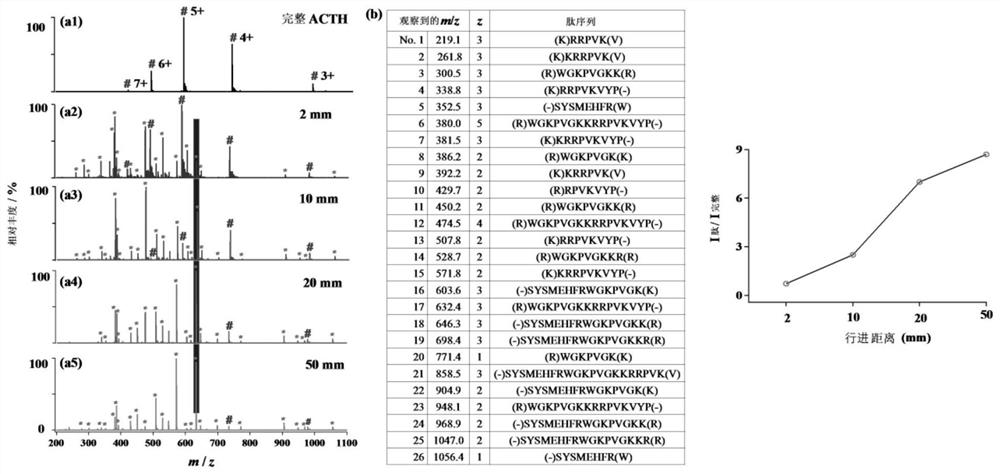
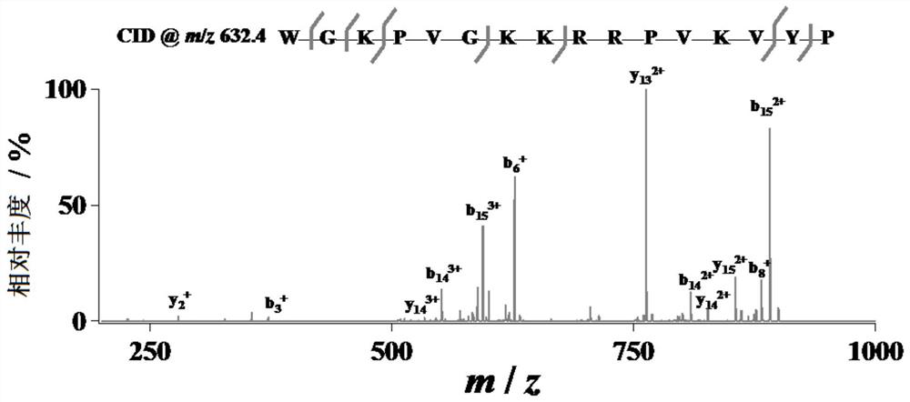
[0086] To optimize the performance of droplet-MS, trypsinized ACTH was used as a simple model system.
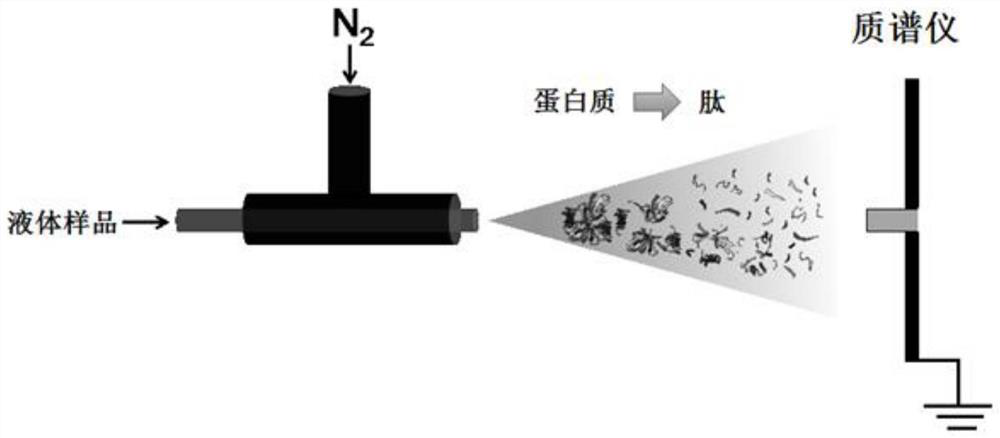
[0087] 5 mM ammonium bicarbonate (NH 4 HCO 3 , pH 8) aqueous solution sample was injected into a self-made nebulizer (the inner diameter of the capillary was 50 μm, the outer diameter was 148 μm), thereby generating a micro-droplet flow (such as figure 1 shown).
[0088] Dry N at a pressure of 120 psi 2 With the aid of nebulizing gas, the sample solution was sprayed from the tip of a fused quartz capillary (outer diameter 148 μm, inner diameter 50 μm, Polymicro Technologies, China). Droplets were directed into MS for real-time detection by placing the nebulizer at an appropriate location in front of a high-resolution mass spectrometer (LTQ Orbitrap Elite, Thermo Scientific, San Jose, CA). The MS inlet capillary was kept at 275°C and the capillary voltage was kept at 0V. No other source of gas is used when digestion is perform...
Embodiment 2
[0099] Digestion and analysis of proteins that are particularly difficult to digest with trypsin using Microdroplet-MS.
[0100] Add myoglobin (10 μM) and trypsin (5 μg / mL) in 5 mM ammonium bicarbonate (NH 4 HCO 3 , pH 8) aqueous solution sample was injected into a self-made nebulizer (the inner diameter of the capillary was 50 μm, the outer diameter was 148 μm), thereby generating a micro-droplet flow (such as figure 1 shown).
[0101] Dry N at a pressure of 120 psi 2 With the aid of nebulizing gas, the sample solution was sprayed from the tip of a fused quartz capillary (outer diameter 148 μm, inner diameter 50 μm, Polymicro Technologies, China). Droplets were introduced into the MS by placing the nebulizer in an appropriate position in front of a high-resolution mass spectrometer (LTQ Orbitrap Elite, Thermo Scientific, San Jose, CA) and applying a +3 kV positive high voltage (BOHER HV, Genvolt, UK) to the nebulizer. Perform real-time detection. The MS inlet capillary w...
Embodiment 3
[0113] Digestion and analysis of cytochrome c by microdroplet-MS.
[0114] Following the same method as described in Example 1 and Example 2, the droplet-MS was further used for the digestion and analysis of cytochrome c at a positive voltage of +3 kV.
[0115] Such as Figure 8 As shown, 33 peptides of cytochrome c were successfully identified (corresponding to 83% sequence coverage). The results demonstrate that droplet-MS is a versatile tool for protein digestion.
PUM
| Property | Measurement | Unit |
|---|---|---|
| diameter | aaaaa | aaaaa |
Abstract
Description
Claims
Application Information
 - R&D
- Intellectual Property
- Life Sciences
- Materials
- Tech Scout
- Unparalleled Data Quality
- Higher Quality Content
- 60% Fewer Hallucinations
Browse by: Latest US Patents, China's latest patents, Technical Efficacy Thesaurus, Application Domain, Technology Topic, Popular Technical Reports.
© 2025 PatSnap. All rights reserved.Legal|Privacy policy|Modern Slavery Act Transparency Statement|Sitemap|About US| Contact US: help@patsnap.com


