A fundus image stitching method based on deep neural network

A deep neural network and fundus image technology, applied in the field of medical image processing, can solve the problems of affecting the stitching effect, too few feature points, and unable to verify the stitching results, etc., to achieve accurate stitching images, ensure accuracy, and high stitching efficiency.

Active Publication Date: 2022-05-17
SICHUAN UNIV
View PDF9 Cites 0 Cited by
  • Summary
  • Abstract
  • Description
  • Claims
  • Application Information

AI Technical Summary

Problems solved by technology

[0003] The existing fundus image stitching technology mainly has the following disadvantages: first, too few feature points lead to registration failure, or too many mismatch points lead to wrong matching parameters; second, the stitching scheme cannot be adjusted according to the type of eye disease, which affects Splicing effect; third, the splicing speed is slow, and the splicing results cannot be verified

Method used


Image

  • A fundus image stitching method based on deep neural network
  • A fundus image stitching method based on deep neural network
  • A fundus image stitching method based on deep neural network

Examples


Embodiment Construction

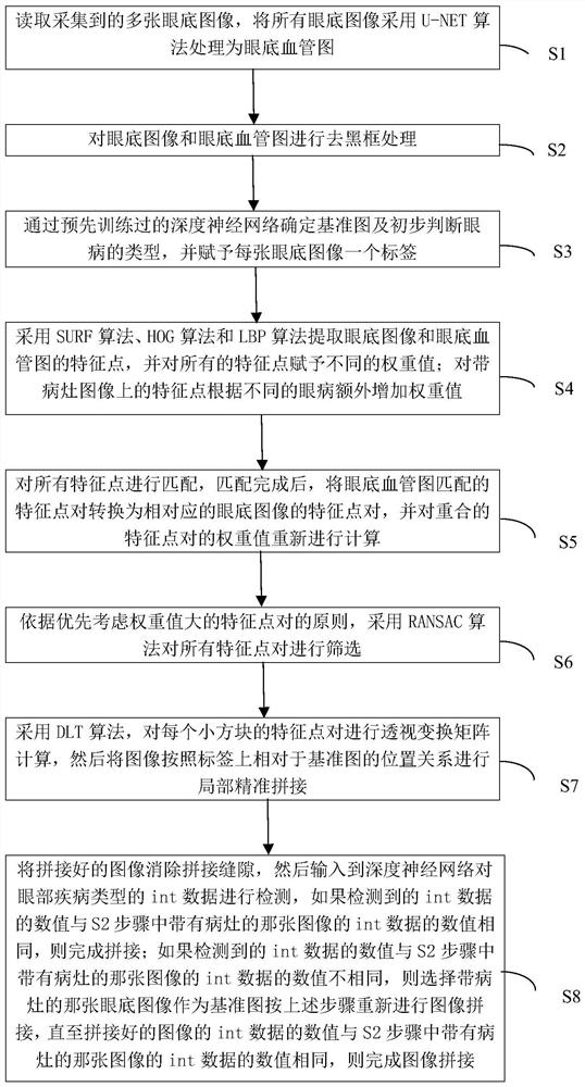
[0028] Such as Figure 1 to Figure 6 As shown, the present embodiment provides a fundus image mosaic method based on a deep neural network comprising the following steps:

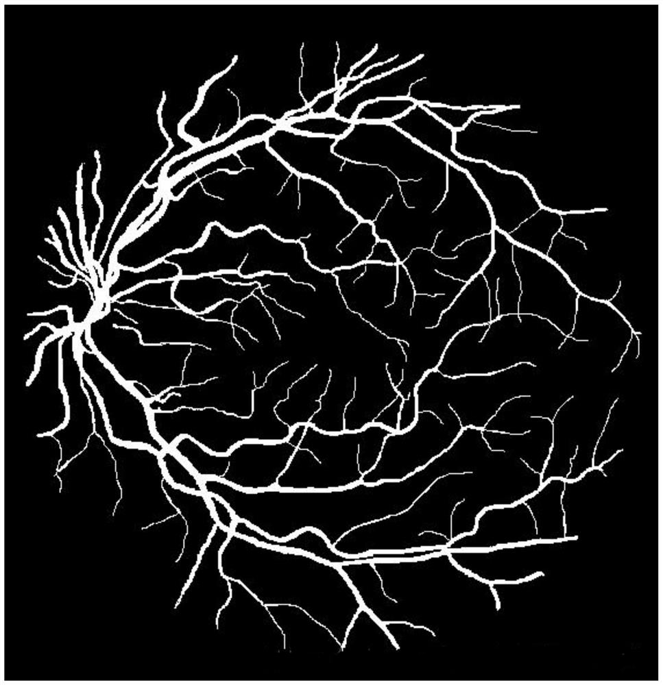
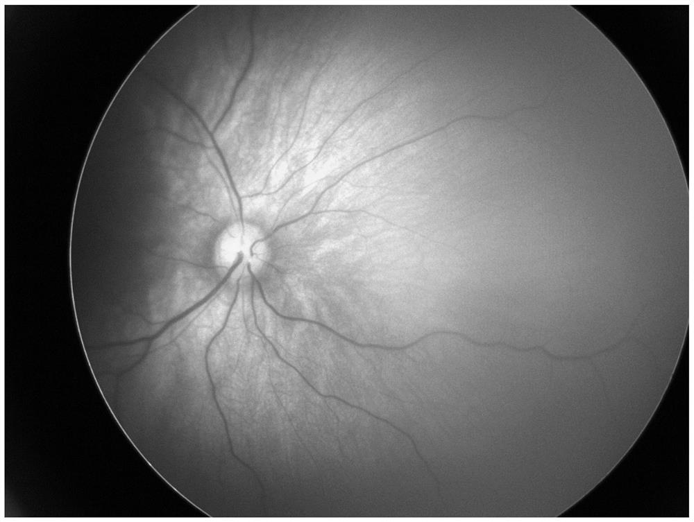
[0029] S1: Read multiple fundus images collected, and process all fundus images into figure 2 The fundus vascular map is shown, and the fundus image and the fundus vascular map are stored in different storage spaces;

[0030] S2: Perform black frame removal processing on the fundus image and fundus blood vessel map. The specific method is to detect each row of the image matrix and remove all pixels with a value of zero;

[0031] S3: Determine the reference map and preliminarily judge the type of eye disease through the pre-trained deep neural network, and assign a label to each fundus image, which records whether the fundus image is a reference map and the position of the fundus image relative to the reference map , and whether the fundus image expressed by int data has lesions and eye disease types;

...


PUM


Abstract

The invention discloses a method for mosaicing fundus images based on a deep neural network, comprising the following steps: S1 reads a plurality of collected fundus images, and processes the fundus images into a fundus blood vessel map; S2 removes the fundus image and the fundus blood vessel map Black frame processing; S3 determines the reference map and preliminarily judges the type of eye disease through the pre-trained deep neural network, and assigns a label to each fundus image; S4 uses SURF algorithm, HOG algorithm and LBP algorithm to extract the fundus image and fundus blood vessel map feature points, and assign different weight values ​​to all feature points; S5 matches all feature points; S6 uses the RANSAC algorithm to screen all feature point pairs according to the principle of preferentially retaining feature point pairs with large weight values; S7 calculates feature points The perspective transformation matrix of point pairs is used to stitch the images; S8 inputs the stitched images to the deep neural network for detection. The invention can improve the accuracy and splicing efficiency of spliced ​​images.

Description

technical field [0001] The invention relates to a fundus image mosaic method based on a deep neural network, belonging to the technical field of medical image processing. Background technique [0002] At present, fundus images are generally obtained by fundus cameras. Due to the limitations of fundus cameras, the obtained images can only be partial images of the fundus, which makes ophthalmologists only rely on naked eye observation and manual alignment in clinical diagnosis and treatment. , not only the efficiency is low, but also the accuracy cannot be guaranteed. There are two ways to solve this problem. One is to increase the field of view of equipment imaging, but this usually requires relatively expensive expenses and is unrealistic for most hospitals. Another approach is to splice multiple fundus images so that the entire fundus image of the patient can be presented on one image, so as to meet the needs of clinical diagnosis and treatment. [0003] The existing fund...

Claims


Application Information

Patent Timeline
Patent Type & Authority Patents(China)
IPC IPC(8): G06T3/40G06T7/00G06V40/18G06V10/50G06V10/46G06V10/82G06N3/04
CPCG06T3/4038G06T7/0012G06T2207/20021G06T2207/30041G06N3/045
Inventor 邹耀徵龚炜文一帆文怀敏付源溟王沐珊王秋昊李鑫宇
Owner SICHUAN UNIV
Why Eureka
Try Eureka