
Cancer auxiliary diagnosis device and method based on digital pathological image

A technology for digital pathology and auxiliary diagnosis, applied in medical equipment, medical automated diagnosis, computer-aided medical procedures, etc., can solve problems such as time-consuming diagnosis process, inability to share case diagnosis results with clients, inability to compare and analyze digital pathology images, etc. , to achieve the effect of shortening the time

Pending Publication Date: 2021-10-29
冯浩
View PDF0 Cites 0 Cited by
  • Summary
  • Abstract
  • Description
  • Claims
  • Application Information

AI Technical Summary

Problems solved by technology

[0004] Aiming at the deficiencies of the prior art, the present invention provides a cancer auxiliary diagnosis device and diagnosis method based on digital pathological images, which solves the problem that the existing cancer auxiliary diagnosis The comparative analysis between the digital pathology images and the existing cases in the database results in a large time-consuming diagnosis process, and it is impossible to quickly share the diagnosis results of the cases with other clients, and the auxiliary cancer diagnosis equipment is not convenient for pathological slices. After fixing and uninstalling, the problem that partial pathological images cannot be enlarged and displayed separately

Method used


Image

  • Cancer auxiliary diagnosis device and method based on digital pathological image
  • Cancer auxiliary diagnosis device and method based on digital pathological image
  • Cancer auxiliary diagnosis device and method based on digital pathological image

Examples


Embodiment Construction

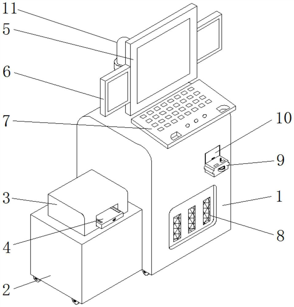
[0049] The following will clearly and completely describe the technical solutions in the embodiments of the present invention with reference to the accompanying drawings in the embodiments of the present invention. Obviously, the described embodiments are only some, not all, embodiments of the present invention.

[0050] In the description of the present invention, unless otherwise stated, the meaning of "plurality" is two or more; the terms "upper", "lower", "left", "right", "inner", "outer" , "front end", "rear end", "head", "tail", etc. indicate the orientation or positional relationship based on the orientation or positional relationship shown in the drawings, and are only for the convenience of describing the present invention and simplifying the description, rather than Nothing indicating or implying that a referenced device or element must have a particular orientation, be constructed, and operate in a particular orientation should therefore not be construed as limiting ...


PUM


Abstract

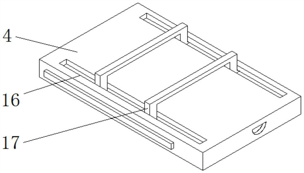
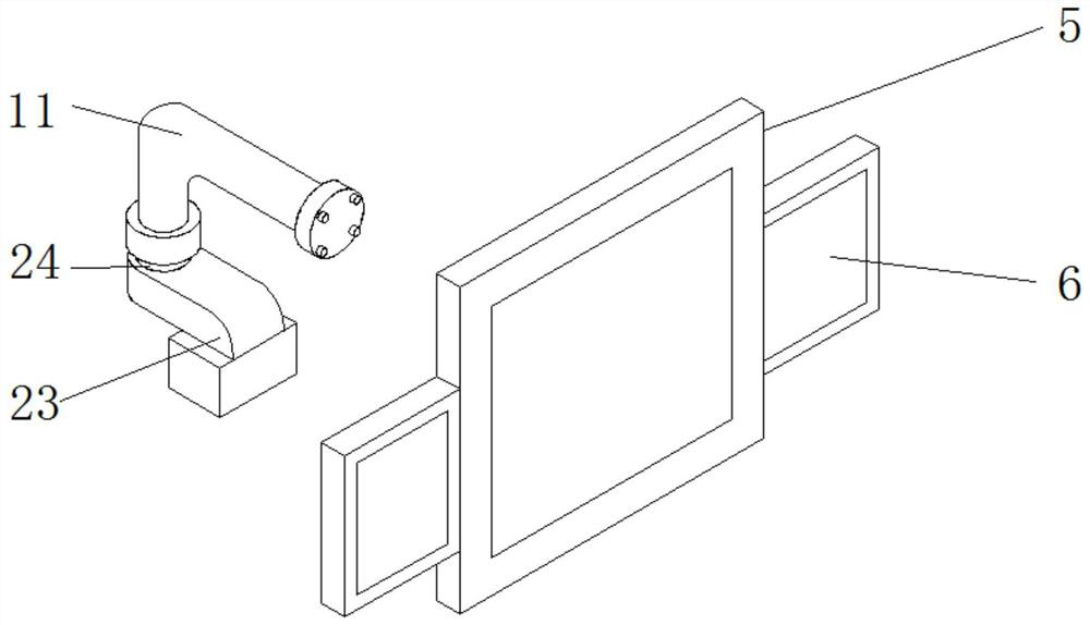
The invention discloses a cancer auxiliary diagnosis device and method based on a digital pathological image, relates to the technical field of cancer auxiliary diagnosis devices, and in particular relates to the cancer auxiliary diagnosis device and method based on the digital pathological image. The cancer auxiliary diagnosis device comprises a device box body, and the left side of the device box body is provided with a supporting box body; a dustproof filter screen is arranged on the outer wall of the device box body; a data comparison module is arranged on the outer wall of a main display screen, and a PLC control module is arranged on the left side of the data comparison module; and a data analysis module is arranged on the right side of a data receiving module, and a data sending module is arranged on the right side of the data analysis module. According to the cancer auxiliary diagnosis device and method based on the digital pathological image, the data comparison module is arranged, the digital pathological image to be diagnosed and case data in an existing database can be compared and analyzed, and therefore auxiliary diagnosis can be conveniently conducted on existing cases according to similar cases.

Description

technical field [0001] The invention relates to the technical field of cancer auxiliary diagnosis equipment, in particular to an auxiliary cancer diagnosis equipment and a diagnosis method based on digital pathological images. Background technique [0002] Digital pathological full slides refer to the use of slice scanning imaging systems to convert pathological slices into high-magnification large-scale digital images that can be displayed, transmitted, and processed by computers. Taking advantage of the characteristics of easy storage, transmission and online viewing of digital pathological slides, the remote diagnosis system of cancer based on pathological images has been developed rapidly. Relying on this cancer remote diagnosis technology, some county-level hospitals in remote areas of the country have already met the conditions for cancer diagnosis. However, according to the statistics of the current remote diagnosis platform, the rate of agreement between the diagnos...

Claims


Application Information

Patent Timeline
Patent Type & Authority Applications(China)
IPC IPC(8): G16H50/20G16H40/63
CPCG16H50/20G16H40/63
Inventor 冯浩李硕杨义李彦生李林起冯潇然陶冶孙晗雅
Owner 冯浩
Learn More