
A medical image recognition method, device, computer equipment and storage medium

A medical image and recognition method technology, applied in the field of medical image processing, can solve the problems of low efficiency of manual recognition and recognition, achieve objective recognition results, improve recognition efficiency, and improve recognition accuracy

Active Publication Date: 2022-01-04
WUHAN ENDOANGEL MEDICAL TECH CO LTD
View PDF7 Cites 0 Cited by
  • Summary
  • Abstract
  • Description
  • Claims
  • Application Information

AI Technical Summary

Problems solved by technology

[0003] The embodiment of the present application provides a medical image recognition method, device, computer equipment and storage medium to solve the technical problem of low recognition efficiency in manual recognition

Method used


Image

  • A medical image recognition method, device, computer equipment and storage medium
  • A medical image recognition method, device, computer equipment and storage medium
  • A medical image recognition method, device, computer equipment and storage medium

Examples


Embodiment Construction

[0038] The following will clearly and completely describe the technical solutions in the embodiments of the present invention with reference to the accompanying drawings in the embodiments of the present invention. Obviously, the described embodiments are only some, not all, embodiments of the present invention. Based on the embodiments of the present invention, all other embodiments obtained by persons of ordinary skill in the art without creative efforts fall within the protection scope of the present invention.

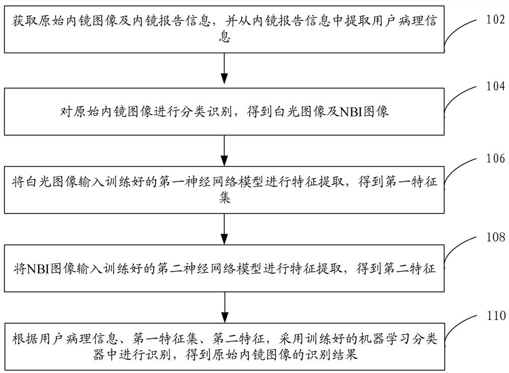
[0039] Such as figure 1 As shown, in one embodiment, a medical image recognition method is provided, and the medical image recognition method can be applied to a terminal or a server, and this embodiment is described by taking the application to a server as an example. The medical image recognition method specifically includes the following steps:

[0040] Step 102, acquiring the original endoscopic image and endoscopic report information, and extracting pathologi...


PUM


Abstract

Embodiments of the present application provide a medical image recognition method, device, computer equipment, and storage medium. The method first acquires the original endoscopic image and endoscopic report information, and extracts pathological information from the endoscopic report information. Then, the original Endoscopic images are classified and recognized to obtain white light images and NBI images. Then, the white light images are input into the trained first neural network model for feature extraction to obtain the first feature set, and the NBI images are input into the trained second neural network. The model performs feature extraction to obtain the second feature. Finally, according to the pathological information, the first feature set, and the second feature, it uses the trained machine learning classifier to perform recognition, and obtains the recognition result of the endoscopic image. The fusion of multiple features of the original endoscopic image improves the recognition accuracy. At the same time, the machine learning classifier is used for recognition, which makes the recognition result more objective and greatly improves the recognition efficiency of medical images.

Description

technical field [0001] The present application relates to the technical field of medical image processing, in particular to a medical image recognition method, device, computer equipment and storage medium. Background technique [0002] Endoscopic image refers to the medical electronic optics that integrates high-tech technologies such as light, machinery, and electricity, which can be inserted into human body cavities and visceral cavities for direct observation, diagnosis, and treatment. The processed image output by the instrument is convenient for doctors to identify and diagnose according to the endoscopic image of the electronic endoscope. However, manual identification has high requirements for endoscopists, which is subjectivity, low efficiency, and professional doctors are required problem of identification. Contents of the invention [0003] Embodiments of the present application provide a medical image recognition method, device, computer equipment, and storage...

Claims


Application Information

Patent Timeline
Patent Type & Authority Patents(China)
IPC IPC(8): G06K9/00G06K9/62G06N3/04G06N3/08
CPCG06N3/08G06N3/045G06F18/24323
Inventor 于红刚卢姿桦姚理文张丽辉
Owner WUHAN ENDOANGEL MEDICAL TECH CO LTD
Learn More