Method for manufacturing human body transparent spinal canal cavity by applying 3D printing technology

A 3D printing and spinal canal technology, which is applied in the field of manufacturing transparent spinal canal cavity of human body, can solve the problems of lack of visual training effect of anesthesia plane

Active Publication Date: 2021-11-26
JILIN UNIV
View PDF18 Cites 0 Cited by
  • Summary
  • Abstract
  • Description
  • Claims
  • Application Information

AI Technical Summary

Problems solved by technology

However, the model lacks an intuitive trai

Method used


Image

  • Method for manufacturing human body transparent spinal canal cavity by applying 3D printing technology
  • Method for manufacturing human body transparent spinal canal cavity by applying 3D printing technology
  • Method for manufacturing human body transparent spinal canal cavity by applying 3D printing technology

Examples


Embodiment Construction

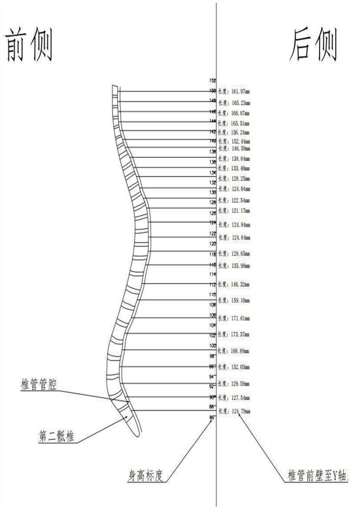
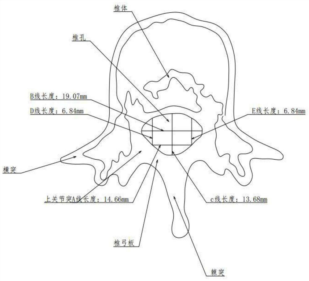
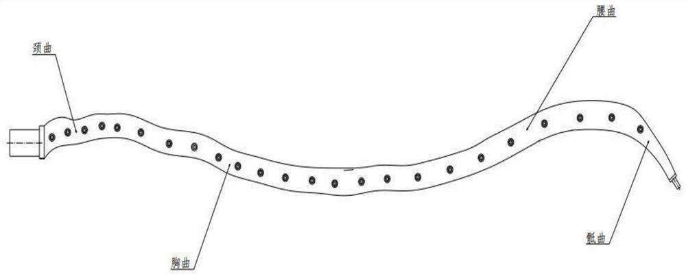
[0024] see Figure 1 to Figure 4 Shown:

[0025] The method of applying 3D printing technology to manufacture the transparent spinal canal cavity of human body provided by the present invention comprises the following steps:

[0026] The first step is to measure and obtain the data of the human spinal canal cavity through computed tomography, that is, CT. The specific steps are as follows:

[0027] Step 1. Curvature of the spinal canal cavity: Obtain the cross-sectional view of the sagittal centerline of the spinal canal in the lateral view of the human body, which is placed vertically in the Cartesian coordinate system, and the Y-axis value represents the height value of each characteristic part of the spinal canal. Y-axis value is accurate to 1mm; X-axis value: establish a horizontal line from the midpoint of the anterior wall of the vertebral canal to the Y-axis, and measure the value as the X-axis value. The X-axis value is accurate to 0.01mm, and each horizontal line is ...


PUM


Abstract

The invention discloses a method for manufacturing a human body transparent spinal canal cavity by applying a 3D printing technology, which comprises the following steps of: 1, measuring and acquiring data of the human body spinal canal cavity through computed tomography, namely CT (Computed Tomography); 2, using CATIA software to generate a 3D image according to the measured data; and 3, inputting the 3D printing data of the vertebral canal cavity into a 3D printer, and generating the bionic human body transparent vertebral canal cavity by applying a transparent polyester material through 3D printing. The method has the beneficial effects that when the bionic human body transparent spinal canal cavity is filled with bionic colorless cerebrospinal fluid, the pharmacokinetic law of anesthetics with obvious colors in the spinal canal cavity can be observed and researched under direct vision, and quantitative research data and technical training of anesthesia plane control are obtained. Quantitative research on subarachnoid space anesthesia level control factors can be carried out, and meanwhile, extremely favorable help can be provided for anesthesia department doctors and medical college school students to carry out anesthesia level control factor training.

Description

technical field [0001] The invention relates to a method for manufacturing a transparent spinal canal cavity of a human body, in particular to a method for manufacturing a transparent spinal canal cavity of a human body by using 3D printing technology. Background technique [0002] Currently, subarachnoid nerve block is an important clinical anesthesia technique. The principle is: puncture in the intervertebral space below the second lumbar vertebra, and after the tip of the puncture needle enters the subarachnoid space, tightly connect the syringe containing the anesthetic solution to the end of the puncture needle, push the syringe, and inject the anesthetic solution into the in the cerebrospinal fluid in the subarachnoid space. The diffusion range of the anesthetic solution in the cerebrospinal fluid (anesthesia plane: the anesthetic solution produces anesthesia on the nerve roots and spinal cord in the soaked range.) is determined by the following factors: injection spe...

Claims


Application Information

Patent Timeline
IPC IPC(8): B29C64/386B33Y50/00A61B5/107A61B6/03
CPCB29C64/386B33Y50/00A61B5/107A61B6/03
Inventor 孙旭芳刘畅赵元愉关慧文
Owner JILIN UNIV
Why Eureka
Try Eureka