Method for detecting lower muscular fascia and included angle between lower muscular fascia and muscle fiber based on B-mode ultrasound image

An image detection and myofascial technology, applied in the field of image processing, can solve problems such as insufficient data, high noise in ultrasonic images, insufficient cognition, etc., and achieve the effect of auxiliary diagnosis

Pending Publication Date: 2021-12-07
杭州类脑科技有限公司
View PDF0 Cites 0 Cited by
  • Summary
  • Abstract
  • Description
  • Claims
  • Application Information

AI Technical Summary

Problems solved by technology

However, due to insufficient data, high noise in ultrasound images, and insufficient

Method used


Image

  • Method for detecting lower muscular fascia and included angle between lower muscular fascia and muscle fiber based on B-mode ultrasound image
  • Method for detecting lower muscular fascia and included angle between lower muscular fascia and muscle fiber based on B-mode ultrasound image
  • Method for detecting lower muscular fascia and included angle between lower muscular fascia and muscle fiber based on B-mode ultrasound image

Examples


Embodiment Construction

[0040] Below in conjunction with accompanying drawing and specific embodiment the present invention is described in further detail:

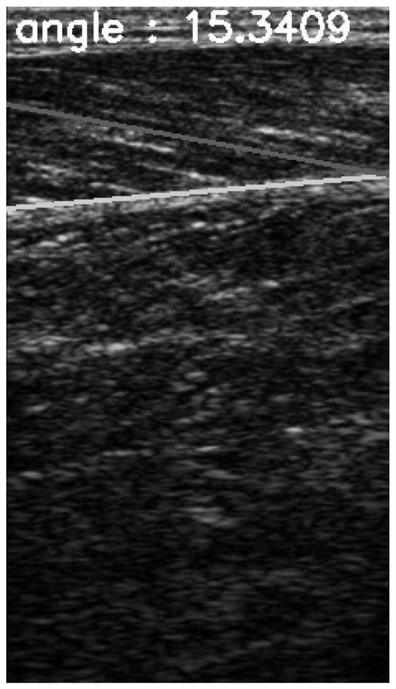
[0041] The method of detecting the position of the lower myofascia and measuring the angle between it and the muscle fiber based on the B-ultrasound image, the specific processing process is as follows:

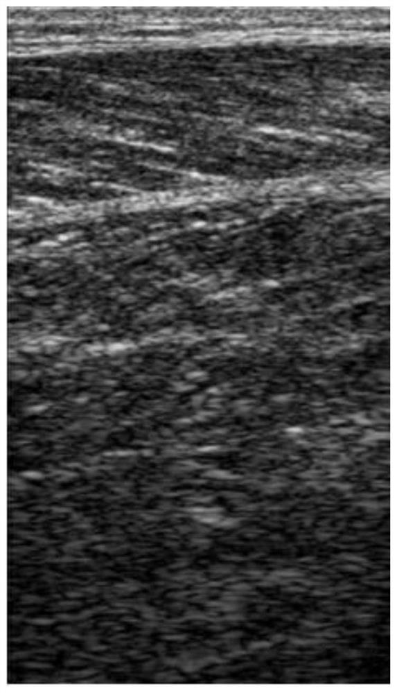
[0042] Process 1: Get such as figure 1 The original B-ultrasound image of the musculoskeletal shown;

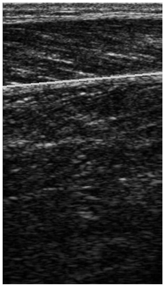
[0043] Process 2: Automatically detect the position of the lower myofascia. The specific processing steps include:

[0044] Step a: Use multi-scale anisotropic filtering to filter and enhance the acquired muscle B-ultrasound image.

[0045] The B-ultrasonic scan image obtained in the note process one is I, wherein any pixel point coordinate is a two-dimensional vector x=(x 1 , x 2 ), where x 1 、x 2 are the components of the vector in the x-direction and y-direction; for any positive number σ>0, define the Gaussian kernel with a kernel wi...


PUM


Abstract

The invention relates to an image processing technology, and aims to provide a method for detecting a lower muscular fascia and an included angle between the lower muscular fascia and a muscle fiber based on a B-mode ultrasound image. The method comprises the following steps: acquiring an original muscle B ultrasonic scanning image; enhancing the acquired muscle B ultrasonic scanning image by using multi-scale anisotropic filtering, and extracting a possible lower muscular fascia position based on the enhanced image; then, detecting the lower muscular fascia position of the original B-mode ultrasound image according to each possible element of the lower muscular fascia position; and automatically detecting the possible position and direction of the muscle fiber, and analyzing the included angle between the muscle fiber and the lower muscular fascia. Compared with the prior art, a doctor can be rapidly and automatically helped to quantitatively judge the included angle of the current muscular fascia, and the doctor is assisted in diagnosing related muscle diseases.

Description

technical field [0001] The invention relates to the technical field of image processing, in particular to a method for detecting inferior myofascia and its angle with muscle fibers based on a B-ultrasound image. Background technique [0002] Ultrasound imaging is a commonly used diagnostic tool in clinical practice. In clinical applications, doctors usually use ultrasound image detection to observe the lower myofascia of important muscles and the shape and angle of muscle fibers. Generally speaking, if a muscle is diseased or strained, the shape of its muscle fibers will change. [0003] However, in clinical practice, it is difficult to distinguish the specific magnitude and scope of this change with the naked eye. However, because ultrasound images are noisy and usually contain a lot of images, manual labeling takes a lot of time and is not repeatable, so it is not suitable for the current actual medical situation in our country. [0004] Therefore, it is of great signif...

Claims


Application Information

Patent Timeline
IPC IPC(8): G06T7/00G06T5/30A61B8/08A61B8/00
CPCG06T7/0012G06T5/30A61B8/08A61B8/5207A61B8/5215G06T2207/10132
Inventor 孔德兴郑哲岚牟芸厉竟罗定存李世岩张燕赵博文王春林陈燕王洪霞包凌云彭成忠
Owner 杭州类脑科技有限公司
Why Eureka
Try Eureka