Method for matching and displaying images of different staining pathological sections

A technology of pathological slices and display methods, which is applied in the field of image matching and display of different stained pathological slices, which can solve the problems of large differences in pathological slices and the inability to realize automatic matching and display of different slice images, so as to improve the reading experience and facilitate observation , Good analysis and diagnosis effect

Pending Publication Date: 2022-03-22
NINGBO SUNNY INSTR
View PDF0 Cites 0 Cited by
  • Summary
  • Abstract
  • Description
  • Claims
  • Application Information

AI Technical Summary

Problems solved by technology

However, due to the large differences in the conditions of different stained pathological sections, it is difficult to manually move to the same area, and the existing pathological digital section display technology cannot automatically match and display images of different sections

Method used


Image

  • Method for matching and displaying images of different staining pathological sections
  • Method for matching and displaying images of different staining pathological sections
  • Method for matching and displaying images of different staining pathological sections

Examples


Embodiment Construction

[0055] In order to more clearly describe the embodiments of the present invention or the technical solutions in the prior art, the following will briefly introduce the drawings that are used in the embodiments. Apparently, the drawings in the following description are only some embodiments of the present invention, and those skilled in the art can also obtain other drawings according to these drawings without creative efforts.

[0056] The present invention will be described in detail below in conjunction with the accompanying drawings and specific embodiments, and the embodiments cannot be repeated here one by one, but the embodiments of the present invention are not therefore limited to the following embodiments.

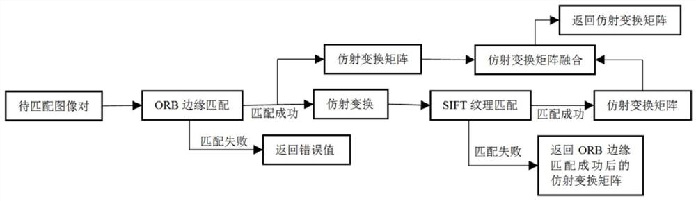
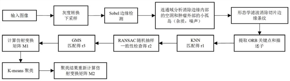
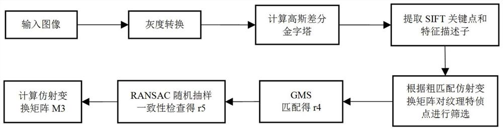
[0057] The method for image matching and displaying of different stained pathological slices of the present invention first stains the continuous slices of the same tissue with different staining reagents, obtains corresponding digital pathological slices through a...


PUM


Abstract

The invention relates to a method for matching and displaying images of different staining pathological sections. The method comprises the following steps: a, dividing a plurality of pathological sections into main sections and auxiliary sections; b, acquiring an affine transformation matrix between the main slice and each auxiliary slice; and c, synchronously displaying the main slice and the auxiliary slice in the main screen and the auxiliary screen respectively. According to the method, a pathologist can accurately observe the same area of the continuous slices, so that the slice reading experience is improved, and the pathologist can better carry out analysis and diagnosis.

Description

technical field [0001] The invention relates to a method for matching and displaying images of different stained pathological slices. Background technique [0002] Pathological examination is an important standard for the diagnosis of malignant tumors. Pathologists mainly make pathological slides of lesion tissues of a certain size according to histopathological methods, and then observe them after generating pathological digital slices through microscopes or pathological scanning equipment. In this way, pathological digital slides gradually become the mainstream reading method for pathological diagnosis, and various pathological digital slice display technologies are becoming more and more mature. In the process of diagnosis of pathological slides, pathologists usually need to continuously slice the same diseased tissue and stain with different reagents (such as HE staining, immunohistochemical IHC staining, etc.), so as to make multiple pathological slides. Therefore, pat...

Claims


Application Information

Patent Timeline
IPC IPC(8): G06T3/00G06T5/00G06T7/12G06T7/13G06T7/187G06T7/40G06V10/46G06V10/762G06F17/16
CPCG06T7/40G06T7/13G06T7/12G06T7/187G06F17/16G06F18/22G06F18/23G06T3/02G06T5/70
Inventor 夏晨洋叶灵枫何英杰
Owner NINGBO SUNNY INSTR
Why Eureka
Try Eureka