An application of promoting atg5-atg12 conjugation to enhance autophagy by scavenging wild-type p53 protein

A technology of ATG5-ATG12 and P53 proteins, applied in the field of biomedicine, can solve the problems of unclear inhibition mechanism and achieve the effect of enhancing the level of autophagy

Active Publication Date: 2022-07-19
ACADEMY OF MILITARY MEDICAL SCI
View PDF5 Cites 0 Cited by
  • Summary
  • Abstract
  • Description
  • Claims
  • Application Information

AI Technical Summary

Problems solved by technology

However, under normal physiological stress conditions, p53 inhibits autophagosome formation, but its specific inhibitory mechanism is unclear

Method used


Image

  • An application of promoting atg5-atg12 conjugation to enhance autophagy by scavenging wild-type p53 protein
  • An application of promoting atg5-atg12 conjugation to enhance autophagy by scavenging wild-type p53 protein
  • An application of promoting atg5-atg12 conjugation to enhance autophagy by scavenging wild-type p53 protein

Examples


Embodiment 1

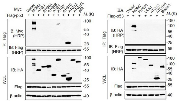
[0027] Example 1 Interaction between p53 and key molecules in the autophagy pathway

[0028] In order to confirm the interaction between p53 and key molecules in the autophagy pathway, HEK293T purchased from ATCC was selected to overexpress the wild-type Flag-p53 plasmid vector and the key molecule expression plasmids in the autophagy pathway (Myc tag and HA tag expression vector plasmids), through The interaction was detected by co-immunoprecipitation technique.

[0029] The implementation steps of the protein co-immunoprecipitation method are as follows:

[0030] 1. Collect cells that have been co-transfected with plasmids for 36 hrs (T25 flask);

[0031] 2. Wash twice with pre-cooled PBS and centrifuge at 3000 rpm for 3 min;

[0032] 3. Add 600 μl of lysis solution (add protease inhibitor and phosphatase inhibitor to the lysis solution, choose HEPES lysis solution or RIPA lysis solution according to specific experimental needs), lyse on ice for 10 min, and then sonicate t...

Embodiment 2

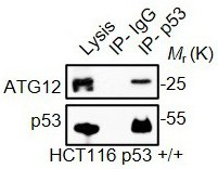
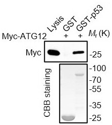
[0050] Example 2 p53 inhibits ATG5-ATG12 conjugation

[0051] After obtaining the interaction between p53 and ATG12, firstly, the wild-type p53 was overexpressed in cells by transfection, and the level of ATG5-ATG12 conjugate was detected. The formation of the yoke body ( Figure 4 ). The specific implementation steps are:

[0052] 1. After 24 hours of cell transfection, blot dry the cell culture medium in the culture plate, and wash twice with PBS with gentle shaking;

[0053] 2. Dry the PBS wash solution, add the cell lysis solution, add an equal volume of 2× loading buffer, mix well to completely lyse the cells and transfer them to an EP tube, boil in boiling water for 15 minutes, and then analyze by SDS-PAGE electrophoresis;

[0054] The ATG5-ATG12 conjugate generation system was further constructed in the extracellular environment, and the purified p53 protein was added. It indicated that wild-type p53 could inhibit the formation of ATG5-ATG12 conjugate by directly bi...

Embodiment 3

[0063] Example 3 p53 inhibits autophagy in mice

[0064] Since the occurrence of autophagy under physiological conditions requires starvation induction, postnatally starved tissue samples were used for detection. In order to verify the effect of p53 on autophagy in vivo, the tissue of neonatal mice starved for 6 hrs with high p53 expression was taken, the tissue cell protein was extracted, and the level of ATG5-ATG12 conjugation was detected. ATG5-ATG12 conjugation levels were significantly reduced ( Figure 7 ); while the level of ATG5-ATG12 conjugation was significantly increased in the tissues of p53 knockout mice ( Figure 8 , Figure 9 ). Figure 9 The scale bar is 50 μm. The specific implementation steps are as follows:

[0065] 1. Preparation and analysis of tissue lysate samples

[0066] 1. Isolate the organs and tissues of newborn mice after starvation;

[0067] 2. Cut 50 mg of tissue, add 500 μl of RIPA lysis solution (add protease inhibitors and phosphatase i...


PUM


Abstract

The invention discloses an application of promoting ATG5-ATG12 conjugation and enhancing cell autophagy by removing p53 protein. First, the mechanism of p53 protein inhibiting cell autophagy is disclosed, and it is found that after p53 protein is increased, the key regulatory protein ATG5 and cell autophagy The conjugation of ATG12 is significantly weakened; by removing p53 protein, the inhibition of p53 protein on ATG5-ATG12 conjugation is reduced, thereby restoring the process of autophagy; the present invention proves that the mechanism of p53 inhibiting autophagy is that p53 inhibits autophagy by binding to ATG12 The conjugation of ATG12-ATG5 can promote the process of autophagy by removing p53 protein, making the inhibition of p53 expression or the removal of p53 protein a potential drug target for relieving autophagy inhibition and promoting autophagy.

Description

technical field [0001] The invention relates to the field of biomedicine, in particular to an application of promoting ATG5-ATG12 conjugation and enhancing cell autophagy by removing p53 protein. Background technique [0002] Autophagy is an important intracellular process, which is highly conserved in the evolution of eukaryotes and can perform material turnover by degrading the components of the cell itself. In addition to normal nutrient metabolism, autophagy can also phagocytose some damaged organelles or proteins in cells into autophagic vesicles with double-membrane structure, and then fuse with lysosomes (animals) to form autophagolysosomes. Remove damaged organelles or proteins from cells. At present, it has been found that autophagy plays an important role in the body's adaptation to starvation, immune response to infection, prevention and treatment of tumors and neurodegenerative diseases, and the deletion or mutation of key genes of autophagy can cause newborn mi...

Claims


Application Information

Patent Timeline
Patent Type & Authority Patents(China)
IPC IPC(8): C12N15/12A01K67/027
CPCC07K14/4746A01K67/0276A01K2227/105A01K2267/03
Inventor 张令强李洪昌李超男崔春萍翟文静
Owner ACADEMY OF MILITARY MEDICAL SCI
Why Eureka
Try Eureka